

Wet but not slippery: Boundary friction in tree frog adhesive toe pads
- PMID: 16971337
- PMCID: PMC1664653
- DOI: 10.1098/rsif.2006.0135
Wet but not slippery: Boundary friction in tree frog adhesive toe pads
Abstract
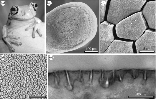
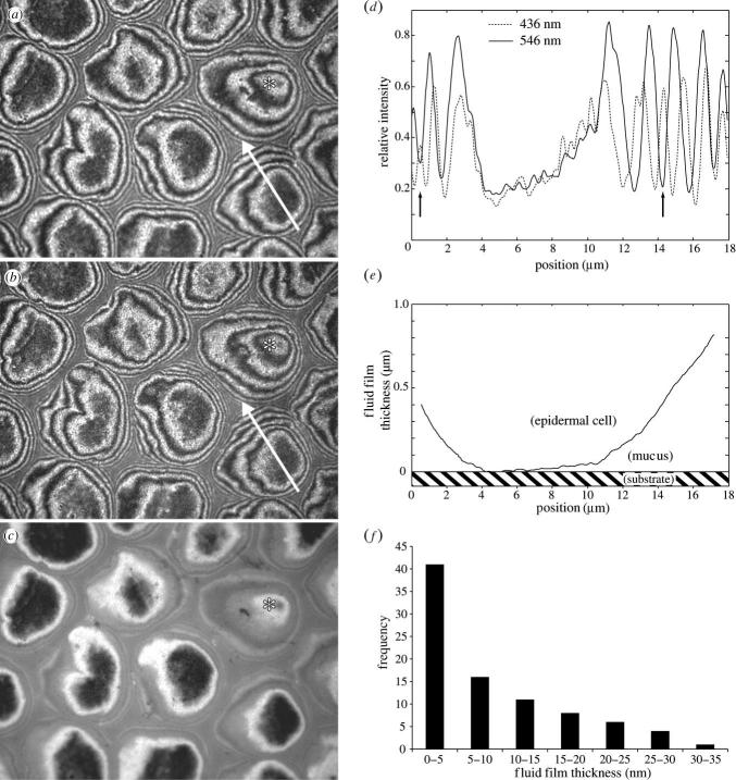
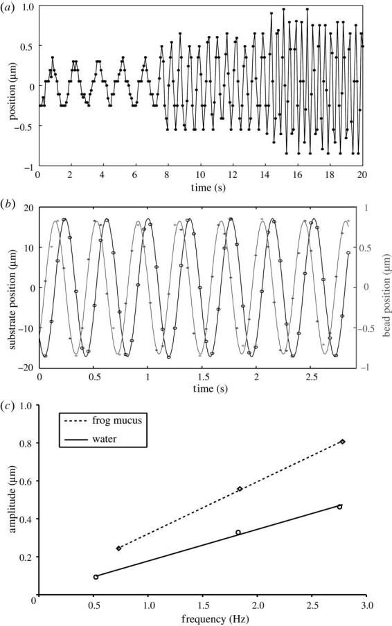
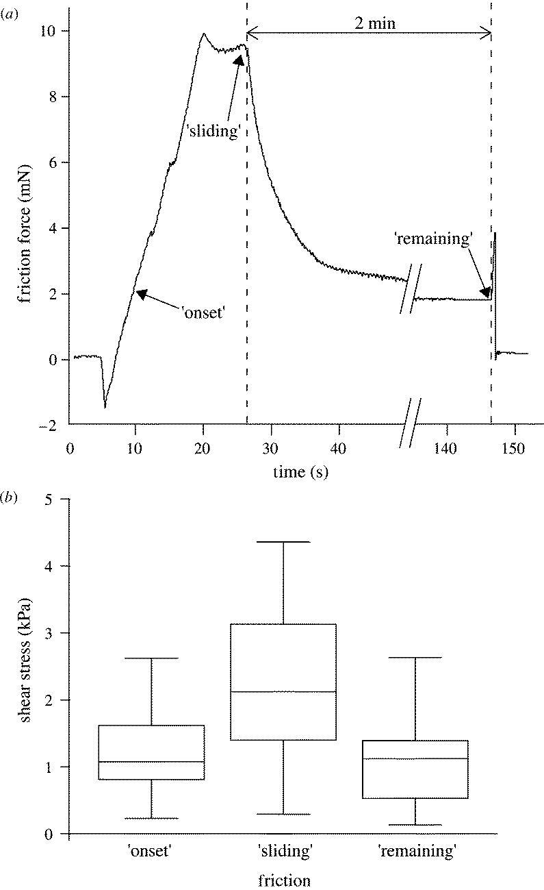
Tree frogs are remarkable for their capacity to cling to smooth surfaces using large toe pads. The adhesive skin of tree frog toe pads is characterized by peg-studded hexagonal cells separated by deep channels into which mucus glands open. The pads are completely wetted with watery mucus, which led previous authors to suggest that attachment is solely due to capillary and viscous forces generated by the fluid-filled joint between the pad and the substrate. Here, we present evidence from single-toe force measurements, laser tweezer microrheometry of pad mucus and interference reflection microscopy of the contact zone in Litoria caerulea, that tree frog attachment forces are significantly enhanced by close contacts and boundary friction between the pad epidermis and the substrate, facilitated by the highly regular pad microstructure.
Figures
Similar articles
-
Nanoscale friction and adhesion of tree frog toe pads.Bioinspir Biomim. 2016 May 11;11(3):035003. doi: 10.1088/1748-3190/11/3/035003. Bioinspir Biomim. 2016. PMID: 27165465
-
Self-cleaning in tree frog toe pads; a mechanism for recovering from contamination without the need for grooming.J Exp Biol. 2012 Nov 15;215(Pt 22):3965-72. doi: 10.1242/jeb.073809. J Exp Biol. 2012. PMID: 23100487
-
Ultrastructure and physical properties of an adhesive surface, the toe pad epithelium of the tree frog, Litoria caerulea White.J Exp Biol. 2009 Jan;212(Pt 2):155-62. doi: 10.1242/jeb.019232. J Exp Biol. 2009. PMID: 19112133 Free PMC article.
-
Tree frog attachment: mechanisms, challenges, and perspectives.Front Zool. 2018 Aug 23;15:32. doi: 10.1186/s12983-018-0273-x. eCollection 2018. Front Zool. 2018. PMID: 30154908 Free PMC article. Review.
-
Tree frog adhesion biomimetics: opportunities for the development of new, smart adhesives that adhere under wet conditions.Philos Trans A Math Phys Eng Sci. 2019 Jul 29;377(2150):20190131. doi: 10.1098/rsta.2019.0131. Epub 2019 Jun 10. Philos Trans A Math Phys Eng Sci. 2019. PMID: 31177956 Free PMC article. Review.
Cited by
-
How do the substrate reaction forces acting on a gecko's limbs respond to inclines?Naturwissenschaften. 2015 Feb;102(1-2):1259. doi: 10.1007/s00114-015-1259-6. Epub 2015 Feb 3. Naturwissenschaften. 2015. PMID: 25645733
-
Morphological studies of the toe pads of the rock frog, Staurois parvus (family: Ranidae) and their relevance to the development of new biomimetically inspired reversible adhesives.Interface Focus. 2015 Feb 6;5(1):20140036. doi: 10.1098/rsfs.2014.0036. Interface Focus. 2015. PMID: 25657830 Free PMC article.
-
Vascular and Osteological Morphology of Expanded Digit Tips Suggests Specialization in the Wandering Salamander (Aneides vagrans).J Morphol. 2025 Jan;286(1):e70026. doi: 10.1002/jmor.70026. J Morphol. 2025. PMID: 39780375 Free PMC article.
-
Influencing the adhesion properties and wettability of mucin protein films by variation of the environmental pH.Sci Rep. 2018 Jun 25;8(1):9660. doi: 10.1038/s41598-018-28047-z. Sci Rep. 2018. PMID: 29942027 Free PMC article.
-
Frictional Properties of Biomimetic Micro-Hexagonal-Textured Surfaces Interacting with Soft Counterfaces under Dry and Wet Conditions.Biomimetics (Basel). 2024 Sep 7;9(9):542. doi: 10.3390/biomimetics9090542. Biomimetics (Basel). 2024. PMID: 39329564 Free PMC article.
References
-
- Arzt E, Gorb S, Spolenak R. From micro to nano contacts in biological attachment devices. Proc. Natl Acad. Sci. 2003;100:10 603–10 606. doi:10.1073/pnas.1534701100 - DOI - PMC - PubMed
-
- Autumn K, et al. Evidence for van der Waals adhesion in gecko setae. Proc. Natl Acad. Sci. 2002;99:12 252–12 256. doi:10.1073/pnas.192252799 - DOI - PMC - PubMed
-
- Aveyard R, Binks B.P, Cho W.G, Fisher L.R, Fletcher P.D.I, Klinkhammer F. Investigation of the force-distance relationship for a small liquid drop approaching a liquid–liquid interface. Langmuir. 1996;12:6561–6569. doi:10.1021/la9607868 - DOI
-
- Baldock R.A, Poole I. Video camera calibration for optical densitometry. J. Microsc. 1993;172:49–54. - PubMed
-
- Barnes W.J.P. Tree frogs and tire technology. Tire Technol. Int. 1999;99:42–47.
Publication types
MeSH terms
Grants and funding
LinkOut - more resources
Full Text Sources
Other Literature Sources
