. 2006 Dec;80(23):11556-70.
doi: 10.1128/JVI.01536-06. Epub 2006 Sep 13.

Structurally mapping the diverse phenotype of adeno-associated virus serotype 4

Affiliations

Structurally mapping the diverse phenotype of adeno-associated virus serotype 4

Lakshmanan Govindasamy et al. J Virol. 2006 Dec.

Abstract

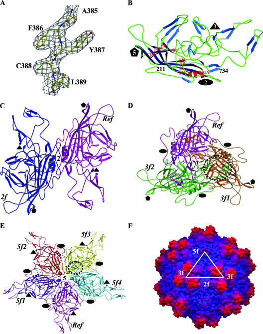
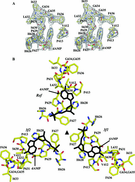
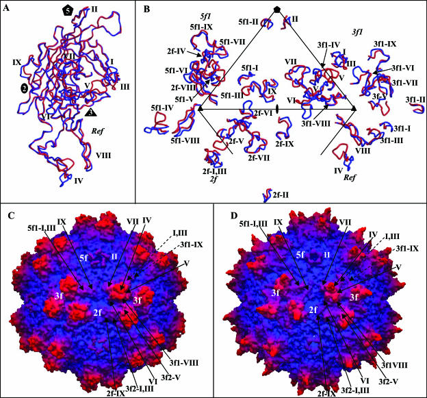
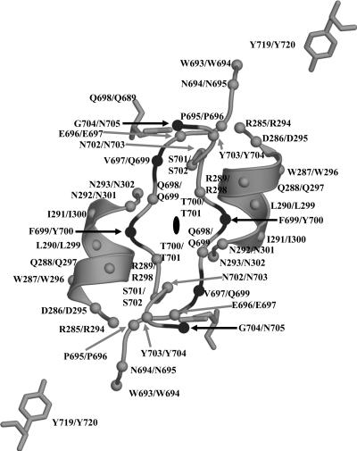
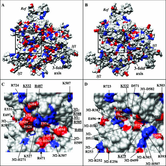
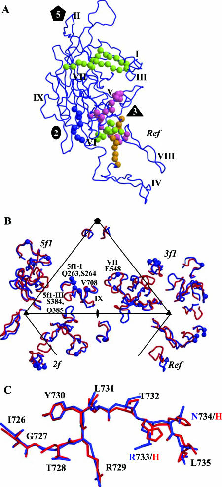
The adeno-associated viruses (AAVs) can package and deliver foreign DNA into cells for corrective gene delivery applications. The AAV serotypes have distinct cell binding, transduction, and antigenic characteristics that have been shown to be dictated by the capsid viral protein (VP) sequence. To understand the contribution of capsid structure to these properties, we have determined the crystal structure of AAV serotype 4 (AAV4), one of the most diverse serotypes with respect to capsid protein sequence and antigenic reactivity. Structural comparison of AAV4 to AAV2 shows conservation of the core beta strands (betaB to betaI) and helical (alphaA) secondary structure elements, which also exist in all other known parvovirus structures. However, surface loop variations (I to IX), some containing compensating structural insertions and deletions in adjacent regions, result in local topological differences on the capsid surface. These include AAV4 having a deeper twofold depression, wider and rounder protrusions surrounding the threefold axes, and a different topology at the top of the fivefold channel from that of AAV2. Also, the previously observed "valleys" between the threefold protrusions, containing AAV2's heparin binding residues, are narrower in AAV4. The observed differences in loop topologies at subunit interfaces are consistent with the inability of AAV2 and AAV4 VPs to combine for mosaic capsid formation in efforts to engineer novel tropisms. Significantly, all of the surface loop variations are associated with amino acids reported to affect receptor recognition, transduction, and anticapsid antibody reactivity for AAV2. This observation suggests that these capsid regions may also play similar roles in the other AAV serotypes.


Figures

FIG. 1.
FIG. 1.
Structure of AAV4. (A) Part of the 2Fo-Fc electron density map of AAV4 (3.2-Å resolution) (gray mesh) for residues 385 to 389, contoured at 1.5σ. (B) Ribbon diagram of AAV4 VP3 showing the core eight-stranded β-barrel strands (dark blue), stretches of antiparallel β-strands (light blue), loops (green), and the helical region (red). The first N-terminal residue observed (211), the C-terminal residue (734), the eight strands (βB to βI) that make up the core β-barrel, and a conserved alpha-helix, αA, are labeled. The secondary structure elements were assigned using the DSSP program (30). (C to E) Ribbon diagrams showing interactions of the AAV4 VP3 monomers at the two-, three-, and fivefold axes, respectively. The reference (Ref), twofold (2f), threefold (3f1 and 3f2), and fivefold (5f1 to 5f4) related monomers are labeled. The interdigitation of two VP3 monomers forming the protrusions around the threefold axis is highlighted by a dashed circle in panel D. The loop between βH and βI that contains interactions between adjacent fivefold related monomers is highlighted by a dashed circle in panel E. The approximate positions of the icosahedral two-, three-, and fivefold axes are shown in panels B to E by filled ovals, triangles, and pentagons, respectively. (F) Depth-cued surface representation of the AAV4 capsid crystal structure viewed down the icosahedral twofold axis. A viral asymmetric unit is depicted by a triangle (in white) bounded by two threefold (3f) axes, divided by a line drawn through the twofold (2f) and a fivefold (5f) axis. Panels A to E were generated using the BOBSCRIPT program (15) and rendered with the RASTER3d program (48); panel F was generated using the GRASP program (51).
FIG. 2.
FIG. 2.
Ordered DNA nucleotide within the AAV4 capsid. (A) Stereoview of 2Fo-Fc electron density map (gray mesh) at the interior pocket containing an ordered DNA nucleotide. The amino acids and the modeled nucleotide (dAMP) are colored according to atom type and labeled. (B) Amino acid residues forming the nucleotide binding pocket, plus the nucleotide viewed through (inside to outside) the threefold axis. The amino acids are colored according to atom type, and the dAMP nucleotide is shown in black. The reference (Ref) and the two threefold related monomers (3f1 and 3f2) are labeled. This figure was generated using the BOBSCRIPT program (15) and rendered with the RASTER3d program (48).
FIG. 3.
FIG. 3.
Structural superimposition and alignment of AAV4 and AAV2 VP3 sequences. (A and B) Atomic models of AAV4 (gray) and AAV2 (black) variable loop regions VI and IX superimposed onto the respective pieces of 2Fo-Fc electron density maps of AAV4 (gray mesh). (C) Structural alignment of the ordered regions of AAV4 and AAV2 VP3 proteins. The alignment was generated by superimposition of the C-α positions of the VP3 atomic models of AAV4 (residues 211 to 734 [VP1 numbering]) and AAV2 (residues 217 to 735 [VP1 numbering]) (PDB accession no. 1LP3). Identical residues and structural regions are shown as white characters in black boxes, superimposable nonidentical residues are shown in black, and regions that adopt a different conformation (nonsuperimposable, with C-α differences of >1.0 Å) are shown in black above (AAV2) and below (AAV4) the aligned residues. The variable regions are labeled I to IX based on the comparison of AAV4 and AAV2 alone and are thus refined from the original definition of Padron et al. (53).
FIG. 4.
FIG. 4.
Comparison of AAV4 and AAV2. (A) Superimposition of coil representations of atomic models of AAV4 (in red) and AAV2 (in blue) (PDB accession no. 1LP3) VP3 monomers. The positions of variable regions I to IX, defined in Fig. 3, are labeled. The approximate two-, three-, and fivefold axes are indicated as in Fig. 1. (B) Positions of variable loop regions I to IX within a viral asymmetric unit. The asymmetric unit is defined in the legend to Fig. 1. The viral asymmetric unit contains contributions from the reference and a twofold (2f), a threefold (3f1), and a fivefold (5f1) VP3 monomer. The prefixes on the labels for the variable regions indicate the contributing monomers. (C and D) Depth-cued surface representations of the AAV4 and AAV2 capsid crystal structures, respectively, viewed down the icosahedral twofold axis. The clustered locations of variable regions I to IX are shown for a viral asymmetric unit of each virus and some of the adjacent regions; the dashed arrows indicate variable regions of the reference outside the asymmetric unit. Panels A and B were generated using the BOBSCRIPT program (15) and rendered with the RASTER3d program (48); panels C and D were generated using the GRASP program (51).
FIG. 5.
FIG. 5.
AAV4 twofold interface. The figure shows a coil representation of the residues lining the depression at the icosahedral twofold axis of the AAV4 capsid and the conserved helix αA on the wall of the depression. The C-α positions are shown as small gray and black balls for amino acids that are conserved and different, respectively, between AAV4 and AAV2. The residue numbers and types are labeled for AAV4/AAV2. These residues form the twofold interface interactions listed in Table 2. The approximate twofold axis is shown as a filled oval. This figure was generated with the BOBSCRIPT program (15) and rendered with the RASTER3d program (48).
FIG. 6.
FIG. 6.
Charge distributions on the AAV2 and AAV4 capsid surfaces. (A and B) Distributions of basic (blue) and acidic (red) residues on theVP3 trimers (Ref, 3f1, and 3f2) of AAV2 and AAV4, respectively, viewed down the icosahedral threefold axis. The approximate twofold axis is indicated with a filled oval. (C and D) Close-up views of the distributions of basic and acidic residues in the AAV2 capsid region (boxed in panel A) mapped as the heparin binding site by mutagenesis (35, 52) and the structurally equivalent region in AAV4 (boxed in panel B), respectively. The residue positions are either labeled on top of the respective surface region or indicated by black arrows. The underlined residues were shown to affect heparin binding in AAV2. This figure was generated using the Chimera program (54).
FIG. 7.
FIG. 7.
Antigenic variation between AAV4 and AAV2. (A) The conformational antigenic sites defined for AAV2 using peptide mapping are shown on a VP3 monomer (blue coil). The C-α positions for the A20 epitope (VP1 numbering; residues 272 to 281, 369 to 378, and 566 to 575) are shown in lime green balls, the C37-B epitope (residues 493 to 502 and 602 to 610) is shown in magenta, the D3 epitope (residues 474 to 483) is shown in orange, and the linear B1 epitope (residues 726 to 733) is shown in blue. The two-, three-, and fivefold axes are depicted as described in the legend to Fig. 1, and variable regions I to IX are labeled. (B) C-α positions (in blue balls) of residues involved in A20 antibody binding and neutralization, as defined by single amino acid mutations mapped onto the AAV4/AAV2 variable regions. The mutated residues and the variable regions in which they are located are labeled. The viral asymmetric unit, as defined in the legend to Fig. 1F, is shown. (C) Superimposition of the AAV2 (blue) and AAV4 (red) structures at the amino acid stretch that forms the B1 epitope. The two amino acids that differ in this region are labeled with the same color as that used for the parent virus structure. This figure was generated using the BOBSCRIPT program (15) and rendered with the RASTER3d program (48).

References

    1. Agbandje, M., S. Kajigaya, R. McKenna, N. S. Young, and M. G. Rossmann. 1994. The structure of human parvovirus B19 at 8 Å resolution. Virology 203:106-115. - PubMed
    1. Agbandje-McKenna, M., and M. S. Chapman. 2006. Correlating structure with function in the viral capsid, p. 125-139. In J. R. Kerr, S. F. Cotmore, M. E. Bloom, R. M. Linden, and C. R. Parrish (ed.), Parvoviruses. Edward Arnold, Ltd., New York, N.Y.
    1. Agbandje-McKenna, M., A. L. Llamas-Saiz, F. Wang, P. Tattersall, and M. G. Rossmann. 1998. Functional implications of the structure of the murine parvovirus, minute virus of mice. Structure 6:1369-1381. - PubMed
    1. Bleker, S., F. Sonntag, and J. A. Kleinschmidt. 2005. Mutational analysis of narrow pores at the fivefold symmetry axes of adeno-associated virus type 2 capsids reveals a dual role in genome packaging and activation of phospholipase A2 activity. J. Virol. 79:2528-2540. - PMC - PubMed
    1. Brunger, A. T. 1992. The free R value: a novel statistical quantity for assessing the accuracy of crystal structures. Nature 355:472-474. - PubMed

Publication types

Associated data

LinkOut - more resources