. 2006 Nov;80(22):11235-40.
doi: 10.1128/JVI.01441-06. Epub 2006 Sep 13.

Two modes of herpesvirus trafficking in neurons: membrane acquisition directs motion

Affiliations

Two modes of herpesvirus trafficking in neurons: membrane acquisition directs motion

Sarah E Antinone et al. J Virol. 2006 Nov.

Abstract

Alphaherpesvirus infection of the mammalian nervous system is dependent upon the long-distance intracellular transport of viral particles in axons. How viral particles are effectively trafficked in axons to either sensory ganglia following initial infection or back out to peripheral sites of innervation following reactivation remains unknown. The mechanism of axonal transport has, in part, been obscured by contradictory findings regarding whether capsids are transported in axons in the absence of membrane components or as enveloped virions. By imaging actively translocated viral structural components in living peripheral neurons, we demonstrate that herpesviruses use two distinct pathways to move in axons. Following entry into cells, exposure of the capsid to the cytosol resulted in efficient retrograde transport to the neuronal cell body. In contrast, progeny virus particles moved in the anterograde direction following acquisition of virion envelope proteins and membrane lipids. Retrograde transport was effectively shut down in this membrane-bound state, allowing for efficient delivery of progeny viral particles to the distal axon. Notably, progeny viral particles that lacked a membrane were misdirected back to the cell body. These findings show that cytosolic capsids are trafficked to the neuronal cell body and that viral egress in axons occurs after capsids are enshrouded in a membrane envelope.


Figures

FIG. 1.
FIG. 1.
Fluorescent viruses propagate with wild-type kinetics. Results from a single-step growth assay of three recombinant viruses (RFP-cap, GFP-env, and RFP-cap/GFP-env) are shown. Adherent cells and media were harvested separately at the indicated times, and titers were quantitated by plaque assay. The RFP-cap virus was previously shown to propagate at rates equal to wild-type PRV (11).
FIG. 2.
FIG. 2.
Incorporation of the gD-GFP fusion protein into extracellular viral particles. (A) Western blot of purified extracellular viral particles (RFP-cap/GFP-env virus) probed with an anti-GFP antibody. (B) Imaging of individual fluorescent viral particles released from cells transfected with the RFP-cap/GFP-env infectious clone. RFP and GFP emissions imaged 2 days posttransfection from the same field are shown. Arrowheads indicate gD-GFP emissions originating from RFP-capsid fusions. The apparent sizes of fluorescent particles are proportional to the brightness of the emissions and not the physical size of the viral particles, which are smaller than the spatial resolution. The field is 19.2 μm by 14.9 μm. (C) Histogram of gD-GFP emissions from dual-fluorescent viral particles as imaged in panel B. GFP emissions were normalized to RFP emissions to correct for variances in the light path. Particles that lacked detectable GFP emissions were not included in the data set.
FIG. 3.
FIG. 3.
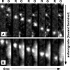
Capsids transport with gD in the anterograde direction only. (A to C) Example time-lapse series of alternating RFP (R) and GFP (G) image captures are shown from left to right following infection of peripheral neurons with the RFP-cap/GFP-env virus. Frames were captured in an axon of a sensory neuron within the first hour postinfection, when capsids transport to the cell body (A), or following replication in either sensory neurons (B) or sympathetic neurons (C), when progeny capsids transport to the distal axon. A single RFP-capsid is seen moving across the field in each montage. Frames are 2.5 μm by 9.0 μm (A) or 1.3 μm by 5.9 μm (B and C) and were captured continuously using 50-ms exposures. (D) Summary of capsid transport in axons of dorsal root sensory neurons as illustrated in panels A and B. The fraction of capsids transported with (solid) or without (white) gD is shown as a percentage of the total capsids tracked during each stage of infection.
FIG. 4.
FIG. 4.
Confirmation of the anterograde capsid/membrane complex. Transport of an RFP-teg/GFP-env virus particle (A) or an RFP-cap particle labeled with DiOC16 lipophilic dye (B) in axons of DRG sensory neurons at 12 to 15 hpi. Frames of alternating mRFP1 (R) and GFP (G) emissions were captured with continuous 50-ms exposures and are shown from left to right. Frames are 1.2 μm by 7.2 μm (A) or 1.1 μm by 5.3 μm (B).

References

    1. Chada, S. R., and P. J. Hollenbeck. 2003. Mitochondrial movement and positioning in axons: the role of growth factor signaling. J. Exp. Biol. 206:1985-1992. - PubMed
    1. Ch'ng, T. H., and L. W. Enquist. 2005. Neuron-to-cell spread of pseudorabies virus in a compartmented neuronal culture system. J. Virol. 79:10875-10889. - PMC - PubMed
    1. Del Rio, T., T. H. Ch'ng, E. A. Flood, S. P. Gross, and L. W. Enquist. 2005. Heterogeneity of a fluorescent tegument component in single pseudorabies virus virions and enveloped axonal assemblies. J. Virol. 79:3903-3919. - PMC - PubMed
    1. Dohner, K., A. Wolfstein, U. Prank, C. Echeverri, D. Dujardin, R. Vallee, and B. Sodeik. 2002. Function of dynein and dynactin in herpes simplex virus capsid transport. Mol. Biol. Cell 13:2795-2809. - PMC - PubMed
    1. Holland, D. J., M. Miranda-Saksena, R. A. Boadle, P. Armati, and A. L. Cunningham. 1999. Anterograde transport of herpes simplex virus proteins in axons of peripheral human fetal neurons: an immunoelectron microscopy study. J. Virol. 73:8503-8511. - PMC - PubMed

Publication types

Substances

LinkOut - more resources