. 2006 Nov;80(22):11313-21.
doi: 10.1128/JVI.01737-06. Epub 2006 Sep 13.

A high-throughput method for cloning and sequencing human immunodeficiency virus type 1 integration sites

Affiliations

A high-throughput method for cloning and sequencing human immunodeficiency virus type 1 integration sites

Sanggu Kim et al. J Virol. 2006 Nov.

Abstract

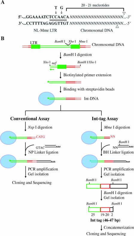
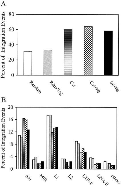
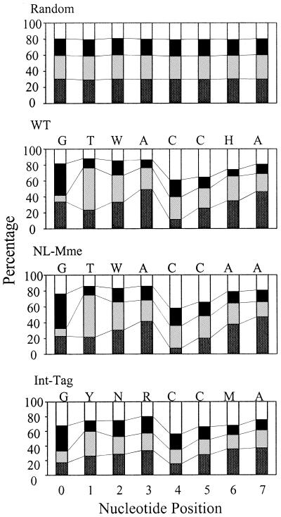
Integration of retroviral DNA is nonspecific and can occur at many sites throughout chromosomes. However, the process is not uniformly distributed, and both hot and cold spots for integration exist. The mechanism that determines target site specificity is not well understood. Because of the nonspecific and widespread nature of integration, studies analyzing the mechanism and factors that control target site selection require the collection and analysis of a large library of human immunodeficiency virus type 1 (HIV-1) proviral clones. Such analyses are time-consuming and labor-intensive using conventional means. We have developed an efficient and high-throughput method of sequencing and mapping a large number of independent integration sites in the absence of any selection or bias. The new assay involves the use of a modified HIV-1 (NL-Mme) containing a type IIS restriction site, MmeI, at the right end of viral DNA. Digestion of genomic DNA from NL-Mme-infected cells generated viral DNA-containing fragments of a discrete size. Subsequent ligation-mediated PCR yielded short integration site fragments termed Int-tags, which were concatemerized for determining multiple integration sites in a single sequencing reaction. Analysis of chromosomal features and sequence preference associated with integration events confirmed the validity of the new high-throughput assay. The assay will aid the effort in understanding the mechanisms of target site selection during HIV-1 DNA integration, and the described methodology can be adapted easily to integration site studies involving other retroviruses and transposons.


Figures

FIG. 1.
FIG. 1.
Assays for genome-wide analysis of HIV-1 integration sites. (A) Construction of mutant HIV with a type IIS MmeI restriction site in the LTR. Bold letters denote viral DNA sequences at the U5 region of the LTR, and italicized letters denote chromosomal DNA. The nucleotides at the positions indicated by the arrows were changed from T and G in the wild-type sequence to C and A, respectively, in the NL-Mme mutant, generating a new recognition site for MmeI (underlined). Arrowheads indicate cleavage sites for MmeI. (B) Schematic diagram outlining the major steps of the conventional assay and the high-throughput Int-tag assay. Viral, cellular, and linker DNAs are denoted by green, red, and black lines or boxes, respectively. Red dotted lines denote cellular DNA with various lengths. Blue ovals represent streptavidin beads, and green diamonds represent biotin. See Materials and Methods for a detailed description of the experimental procedures.
FIG. 2.
FIG. 2.
Replication kinetics of WT and NL-Mme viruses. CEM cells were infected with equal amounts of the p24 equivalent of WT (○) or NL-Mme (□) virus at an MOI of 0.001. The culture media were monitored for p24 levels (ng/ml) at the indicated time points postinfection.
FIG. 3.
FIG. 3.
Distribution of WT and NL-Mme virus integration events in human chromosomes. Results are expressed as the percentages of integration events in each chromosome. Human chromosome numbers are indicated at the bottom of the figure. The numbers of integration events for the random control (open bars), WT virus (gray bars), and NL-Mme virus (black bars) were 5,000, 309, and 323, respectively.
FIG. 4.
FIG. 4.
Analysis by the conventional assay of chromosomal features associated with WT and NL-Mme virus integration events. CEM cells were infected with WT or NL-Mme virus at an MOI of 10. Integration sites were mapped using the conventional assay, and chromosomal features associated with WT (gray bars) and NL-Mme (black bars) proviruses were analyzed. The results are expressed as percentages of total integration events and compared with those of the random control (open bars). Chromosomal features analyzed include transcription units (TU), Alu and mammalian interspersed repeat (MIR) of the SINE, L1 and L2 of the LINE, LTR-E, and DNA-E.
FIG. 5.
FIG. 5.
Mapping of short integration sequences to unique locations and those in identifiable repeat elements. Integration site sequences from cells infected with NL-Mme were determined using the conventional assay (Cvt) or the high-throughput assay (Int-tag). Each sequence was mapped to either unique locations (black bars) or identifiable repeat elements (gray bars), and the results are expressed as percentages of total integration site sequences. The results were compared with those obtained from conventional tags (Cvt-tag), generated by taking the first 19 or 20 nucleotides immediately adjacent to the viral DNA of each integration site sequence determined by the conventional assay, or a random library of 19- and 20-bp cellular sequences (Rdm-tag) generated in silico.
FIG. 6.
FIG. 6.
Analysis of chromosomal features associated with integration events by use of the high-throughput Int-tag assay and the conventional assay. Integration site sequences were determined using the conventional assay (striped bars) or the Int-tag assay (black bars) or generated in silico (open bars). Rdm-tag (gray bars) and Cvt-tag (hatched bars) sequences were derived from integration site sequences generated in silico and by the conventional assay, respectively, as described above. The locations of the integration sites were mapped, and chromosomal features in the vicinity of the integration sites were identified. (A) Transcription units. Integration site sequences that mapped to a unique chromosomal location were analyzed and scored as a part of a transcription unit only if the transcription unit was a member of the RefSeq genes. (B) Identifiable repeat sequences. Integration site sequences that mapped to multiple locations were analyzed for identifiable repeat elements, including Alu and mammalian interspersed repeat (MIR) of the SINE, L1 and L2 of the LINE, LTR-E, and DNA-E.
FIG. 7.
FIG. 7.
Base preference in genomic sequence immediately adjacent to integration sites. The numbers on the x axis represent nucleotide positions of human DNA adjacent to the proviral DNA, where the point of joining between the HIV and human DNA lies to the left of position 0. The height of the bar represents the percent frequency of each base. A, T, G, and C are denoted by dark gray, light gray, black, and open bars, respectively. The preferred sequence is listed on the top of each panel. H denotes A, C, or T but not G; M denotes A or C; N denotes A, C, G, or T; R denotes A or G; W denotes A or T; and Y denotes C or T.

Similar articles

Cited by

References

    1. Ausubel, F. A., R. Brent, R. E. Kingston, D. D. Moore, J. G. Seidman, J. A. Smith, and K. Struhl. 1999. Current protocols in molecular biology. Wiley, New York, N.Y.
    1. Balakrishnan, M., and C. B. Jonsson. 1997. Functional identification of nucleotides conferring specificity to retroviral integrase reactions. J. Virol. 71:1025-1035. - PMC - PubMed
    1. Barr, S. D., J. Leipzig, P. Shinn, J. R. Ecker, and F. D. Bushman. 2005. Integration targeting by avian sarcoma-leukosis virus and human immunodeficiency virus in the chicken genome. J. Virol. 79:12035-12044. - PMC - PubMed
    1. Brown, P. O. 1997. Integration, p. 161-204. In J. M. Coffin, S. H. Hughes, and H. E. Varmus (ed.), Retroviruses. Cold Spring Harbor Laboratory Press, Cold Spring Harbor, N.Y.
    1. Bushman, F., M. Lewinski, A. Ciuffi, S. Barr, J. Leipzig, S. Hannenhalli, and C. Hoffmann. 2005. Genome-wide analysis of retroviral DNA integration. Nat. Rev. Microbiol. 3:848-858. - PubMed

Publication types

LinkOut - more resources