. 2006 Nov;80(22):10931-41.
doi: 10.1128/JVI.01287-06. Epub 2006 Sep 13.

Characterization of human metapneumovirus F protein-promoted membrane fusion: critical roles for proteolytic processing and low pH

Affiliations

Characterization of human metapneumovirus F protein-promoted membrane fusion: critical roles for proteolytic processing and low pH

Rachel M Schowalter et al. J Virol. 2006 Nov.

Abstract

Human metapneumovirus (HMPV) is a recently described human pathogen of the pneumovirus subfamily within the paramyxovirus family. HMPV infection is prevalent worldwide and is associated with severe respiratory disease, particularly in infants. The HMPV fusion protein (F) amino acid sequence contains features characteristic of other paramyxovirus F proteins, including a putative cleavage site and potential N-linked glycosylation sites. Propagation of HMPV in cell culture requires exogenous trypsin, which cleaves the F protein, and HMPV, like several other pneumoviruses, is infectious in the absence of its attachment protein (G). However, little is known about HMPV F-promoted fusion, since the HMPV glycoproteins have yet to be analyzed separately from the virus. Using syncytium and luciferase reporter gene fusion assays, we determined the basic requirements for HMPV F protein-promoted fusion in transiently transfected cells. Our data indicate that proteolytic cleavage of the F protein is a stringent requirement for fusion and that the HMPV G protein does not significantly enhance fusion. Unexpectedly, we also found that fusion can be detected only when transfected cells are treated with trypsin and exposed to low pH, indicating that this viral fusion protein may function in a manner unique among the paramyxoviruses. We also analyzed the F protein cleavage site and three potential N-linked glycosylation sites by mutagenesis. Mutations in the cleavage site designed to facilitate endogenous cleavage did so with low efficiency, and our data suggest that all three N-glycosylation sites are utilized and that each affects cleavage and fusion to various degrees.


Figures

FIG. 1.
FIG. 1.
Schematic of the HMPV F protein. The F protein is proteolytically processed into two fragments, F1 and F2, which are disulfide linked. Arrows point to potential sites of N-linked glycosylation. The amino acid sequence upstream of the cleavage site is indicated along with the mutations made to this site. FP, fusion peptide; HRA and HRB, heptad repeats A and B; TM, transmembrane domain; WT, wild type.
FIG. 2.
FIG. 2.
Expression of HMPV F and HMPV G. (A) Biotinylation. Vero cells in 6-cm dishes were transfected with 4 μg pCAGGS-HMPV F or pCAGGS-HMPV G. The cells were metabolically labeled for 3 h with Tran35S-label ± 0.3 μg/ml TPCK trypsin prior to the biotinylation of surface proteins. Fifteen percent of the immunoprecipitated protein represents the “total” protein. The remaining 85% was subjected to pull down with streptavidin and represents the “surface” protein. The samples were resolved on a 15% SDS-polyacrylamide gel and visualized by autoradiography. Each side of the same gel was differentially contrast enhanced. (B) In vitro coupled transcription/translation of HMPV G (“in vitro”; right side) run side by side on a 15% SDS-polyacrylamide gel with HMPV G immunoprecipitated from transfected Vero cells (“in vivo”; left side).
FIG. 3.
FIG. 3.
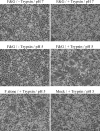
Analysis of HMPV F-promoted cell-cell fusion. (A) Vero cells were transfected with pCAGGS-HMPV F (1 μg) and pCAGGS-HMPV G (1 μg) or the empty pCAGGS vector (1 μg). After 18 to 24 h, confluent cell monolayers were incubated in Opti-MEM ± 0.3 μg/ml TPCK-trypsin for 1 to 2 h and then washed and treated with buffered PBS of the indicated pH for 4 min. The cells were again incubated in Opti-MEM ± trypsin, and the pH pulse was repeated three more times throughout the day before the photographs were taken. (B) Vero cells were transfected with a total of 3.5 μg of DNA consisting of 1.5 μg luciferase cDNA, 1 μg pCAGGS-HMPV F or pCAGGS-SV5 F or empty vector, and 1 μg pCAGGS-HMPV G or pCAGGS-SV5 HN or empty vector. After 18 to 24 h, Vero cells were overlaid on BSR cells expressing the T7 polymerase. The cells were then treated and fusion was analyzed as described in Materials and Methods. The average of the results of four experiments, normalized to the luminosity detected from F-transfected, pH 5-treated cells, is shown. The error bars represent 95% confidence intervals. (C) Six-centimeter dishes of Vero cells were transfected with 1.5 μg luciferase cDNA and 1.5 μg pCAGGS-HMPV F. After 18 to 24 h, Vero cells were overlaid on BSR cells expressing the T7 polymerase. The cells were then treated and fusion was analyzed as described in Materials and Methods. The average of the results of four experiments, normalized to the luminosity of pH 5-treated cells, is shown. The error bars represent 95% confidence intervals. F&G, HMPV F and G proteins present.
FIG. 4.
FIG. 4.
Syncytia promoted by HMPV F in BHK cells. BHK cells were transfected with empty pCAGGS vector (2 μg) or pCAGGS-HMPV F (1 μg) and pCAGGS-HMPV G (1 μg) or empty vector (1 μg). After 18 to 24 h, confluent cell monolayers were incubated in Opti-MEM with 0.2 μg/ml TPCK-trypsin for 1 h, then washed and treated with PBS of the indicated pH for 4 min. The cells were then incubated in DMEM plus FBS for ∼1 h, then in Opti-MEM plus trypsin for ∼1 h. The pH pulse and change of media were repeated three more times before the photographs were taken. F&G, HMPV F and G proteins present.
FIG. 5.
FIG. 5.
Analysis of the surface expression of HMPV F mutants. Vero cells in 6-cm dishes were transfected with 8 μg pCAGGS-HMPV F or a mutant F protein. The cells were metabolically labeled for 3 h with Tran35S-label in the absence (A) or presence (B) of 0.5 μg/ml TPCK trypsin. Biotinylation and analysis were performed as for Fig. 2A, but only the surface populations are shown. Fwt, wild-type F protein.
FIG. 6.
FIG. 6.
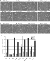
Fusion promoted by HMPV F cleavage-site mutants. Syncytium assays in the presence (A) or absence (B) of trypsin are shown. Vero cells were transfected with 1 μg pCAGGS-HMPV F or a mutant F protein and 1 μg pCAGGS-HMPV G. The cells were treated (each with pH 5 PBS) and analyzed as for Fig. 3A. (C) Reporter gene assay of fusion in the presence of trypsin. Vero cells in 6-cm dishes were transfected with 1.5 μg luciferase cDNA and 1.5 μg pCAGGS-HMPV F or a mutant. After 18 to 24 h, the Vero cells were overlaid on BSR cells expressing the T7 polymerase. The cells were then treated and fusion was analyzed as described in Materials and Methods. The average of the results of three experiments is shown, and the error bars represent 95% confidence intervals. The graph is divided into two sections. The mutants on either side were analyzed on different days but always alongside the wild-type (WT) protein, and values were normalized to that of the wild type in each experiment.
FIG. 7.
FIG. 7.
Analysis of HMPV F N-linked glycosylation. (A) PNGase F treatment of HMPV F and G. Vero cells in 35-mm dishes were transfected with 2 μg pCAGGS-HMPV F, pCAGGS-HMPV G, or empty vector. Cells were metabolically labeled with Tran35S-label for 1 h and chased for 1 h. Immunoprecipitated protein was incubated overnight with or without PNGase F, and samples were resolved on a 15% SDS-polyacrylamide gel and visualized by autoradiography. (B) Vero cells were transfected with 2 μg pCAGGS-HMPV F or a mutant. Cells were metabolically labeled with Tran35S-label for 1 h and chased for 2 h in serum-free DMEM ± 0.3 μg/ml TPCK-trypsin. Immunoprecipitated protein was analyzed as above. (C) Syncytium assay. Vero cells transfected with 1 μg pCAGGS-HMPV F or a mutant and 1 μg pCAGGS-HMPV G were treated (each with pH 5 PBS) and analyzed as for Fig. 3A. (D) Reporter gene assay. Vero cells in 6-cm dishes were transfected with 1.5 μg luciferase cDNA and 1.5 μg pCAGGS-HMPV F or a mutant. After 18 to 24 h, Vero cells were overlaid on BSR cells expressing the T7 polymerase. The cells were then treated and fusion was analyzed as described in Materials and Methods. The average of the results of seven experiments is shown, and the error bars represent 95% confidence intervals.

References

    1. Bagai, S., and R. A. Lamb. 1995. Individual roles of N-linked oligosaccharide chains in intracellular transport of the paramyxovirus SV5 fusion protein. Virology 209:250-256. - PubMed
    1. Bagai, S., and R. A. Lamb. 1995. Quantitative measurement of paramyxovirus fusion: differences in requirements of glycoproteins between simian virus 5 and human parainfluenza virus 3 or Newcastle disease virus. J. Virol. 69:6712-6719. - PMC - PubMed
    1. Barretto, N., L. K. Hallak, and M. E. Peeples. 2003. Neuraminidase treatment of respiratory syncytial virus-infected cells or virions, but not target cells, enhances cell-cell fusion and infection. Virology 313:33-43. - PubMed
    1. Biacchesi, S., Q. N. Pham, M. H. Skiadopoulos, B. R. Murphy, P. L. Collins, and U. J. Buchholz. 2005. Infection of nonhuman primates with recombinant human metapneumovirus lacking the SH, G, or M2-2 protein categorizes each as a nonessential accessory protein and identifies vaccine candidates. J. Virol. 79:12608-12613. - PMC - PubMed
    1. Biacchesi, S., Q. N. Pham, M. H. Skiadopoulos, B. R. Murphy, P. L. Collins, and U. J. Buchholz. 2006. Modification of the trypsin-dependent cleavage activation site of the human metapneumovirus fusion protein to be trypsin independent does not increase replication or spread in rodents or nonhuman primates. J. Virol. 80:5798-5806. - PMC - PubMed

Publication types

LinkOut - more resources