. 2006;34(17):4816-25.
doi: 10.1093/nar/gkl650. Epub 2006 Sep 13.

Amino acid residues critical for RNA-binding in the N-terminal domain of the nucleocapsid protein are essential determinants for the infectivity of coronavirus in cultured cells

Affiliations

Amino acid residues critical for RNA-binding in the N-terminal domain of the nucleocapsid protein are essential determinants for the infectivity of coronavirus in cultured cells

Yong Wah Tan et al. Nucleic Acids Res. 2006.

Abstract

The N-terminal domain of the coronavirus nucleocapsid (N) protein adopts a fold resembling a right hand with a flexible, positively charged beta-hairpin and a hydrophobic palm. This domain was shown to interact with the genomic RNA for coronavirus infectious bronchitis virus (IBV) and severe acute respiratory syndrome coronavirus (SARS-CoV). Based on its 3D structure, we used site-directed mutagenesis to identify residues essential for the RNA-binding activity of the IBV N protein and viral infectivity. Alanine substitution of either Arg-76 or Tyr-94 in the N-terminal domain of IBV N protein led to a significant decrease in its RNA-binding activity and a total loss of the infectivity of the viral RNA to Vero cells. In contrast, mutation of amino acid Gln-74 to an alanine, which does not affect the binding activity of the N-terminal domain, showed minimal, if any, detrimental effect on the infectivity of IBV. This study thus identifies residues critical for RNA binding on the nucleocapsid surface, and presents biochemical and genetic evidence that directly links the RNA binding capacity of the coronavirus N protein to the viral infectivity in cultured cells. This information would be useful in development of preventive and treatment approaches against coronavirus infection.


Figures

Figure 1
Figure 1
Structure of the N-terminal RNA binding domain of IBV N protein. (a) Structure-based alignment of coronavirus nucleocapsid amino acid sequences corresponding to the N-terminal RNA binding domain. Secondary structure elements are labeled above the sequence for IBV-N29–160 and below for the SARS-CoV N-terminal fragment [Huang et al. (18)]. Sequences of IBV (IBV, strain Beaudette, NP_040838); H-CoV (Human Coronavirus, strain HKU1, YP_173242); MHV (Murine Hepatitis Virus, strain 1, AAA46439); TGEV (Porcine Transmissible Gastroenteritis Virus, strain RM4, AAG30228) and SARS (SARS-Coronavirus, 1SSK_A) were obtained from GenBank. Conserved residues are shaded. (b) Surface representation of the IBV-N-terminal fragment with electrostatic potentials colored in blue (positive) and red (negative). Positively charged residues in the flexible hairpin loop and aromatic residues on the hydrophobic floor that are exposed to the solvent and which were subjected to mutagenesis in this study are labeled.
Figure 2
Figure 2
Northwestern analysis of the RNA binding activity of wild-type and mutant N-terminal domain of the IBV N protein. (a) Diagram showing the alanine mutants included in this study. (b) RNA binding assay for the various mutants of the IBV N protein N-terminal domain produced in this study. The binding affinities of mutated IBV N protein N-terminal domains towards positive and negative RNA ligands corresponding to 26 539–27 608 nt of the IBV genome were evaluated by northwestern blot. The mutants were separated by SDS–PAGE, transferred to Hybond C extra membrane and detected by digoxin-labelled RNA. The relative binding affinities, which were assumed to be proportional to the intensities of the detected bands, were normalized with respect to the binding activity of the wild-type domain, which was arbitrarily taken as 100.
Figure 3
Figure 3
Introduction of single amino acid mutations into the IBV genome and recovery of infectious mutant viruses. Vero cells electroporated with the in vitro synthesized transcripts derived from an in vitro assembled full-length clone (rIBV+N), the full-length clone containing the R76A mutation (R76A+N), the full-length clone containing the Y94A mutation (Y94A+N), the full-length clone containing the Q74A mutation (Q74A+N) and the full-length clone containing the Y92A mutation [Y92A+N(1) and (2)] together with the N transcripts. The trans-effects of wild type and mutant N transcripts on rescue of infectious viruses form the in vitro synthesized full-length IBV transcripts were analyzed by electroporation of Vero cells with the in vitro synthesized full-length IBV transcripts together with wild-type (rIBV+N), R76A (rIBV+N-R76A) and Y94 (rIBV+N-Y94A) mutant N transcripts. Cells electroporated with the full-length wild-type IBV transcripts without the N-transcripts (rIBV-N) were included as a negative control. Images were taken at 3 days post-electroporation.
Figure 4
Figure 4
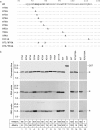
Analysis of negative strand RNA replication and subgenomic RNA transcription in cells transfected with wild-type and mutant full-length transcripts. (a) Detection of subgenomic RNA synthesis in cells electroporated with rIBV (lane 2), R76A (lanes 2–5), Y94A (lanes 6–8), Q74A (lane 9) and Y92A (lane 10) mutant transcripts. Total RNA was prepared from Vero cells electroporated with in vitro synthesized full-length transcripts 3 days post-electroporation. Regions corresponding to the 5′-terminal 415 and 1010 nt of the subgenomic mRNA4 and 3, respectively, were amplified by RT–PCR and analyzed on 1.2% agarose gel. Lane 1 shows DNA markers and numbers on the left indicate the length of DNA in bases. (b) Detection of positive and negative strand RNA synthesis in cells electroporated with rIBV (lanes 8 and 11), Y94A (lanes 3 and 4), R76A (lanes 6 and 7) and Y92A (lanes 9 and 10) mutant transcripts. Total RNA was prepared from Vero cells electroporated with in vitro synthesized full-length transcripts 3 days post-electroporation. Regions corresponding to 14 931–15 600 nt of the positive (+) and negative (−) sense IBV RNA were amplified by RT–PCR and analyzed on 1.2% agarose gel. The negative controls shown in lanes 2 and 5 are PCR analysis of total RNAs extracted from cells transfected with Y94A (lane 2) and R76A (lane 5) using the primer set for negative strand RNA. Lane 1 shows DNA markers. Numbers on the left indicate nucleotides in bases. (c) Quantitative analysis of the negative strand RNA in cells transfected with wild type (lanes 1 and 5), R76A (lanes 2 and 6), Y92A (lanes 3 and 7) and Y94A (lane 4 and 8) by real time RT-PCT at 24 (lane 1–4) and 48 (lanes 5–8) h post-electroporation. The relative ratios of the negative strand RNA in cells transfected with the mutant transcripts to those in cells transfected with wild type transcripts are shown. Numbers on the left indicate nucleotides in bases.
Figure 5
Figure 5
Analysis of the growth properties of wild type and Q74A mutant virus. (a) Plague sizes and one-step growth curves of rIBV and Q74A mutant viruses. Monolayers of Vero cells on a 6-well plate were infected with 100 µl of 1-, 10- and 100-fold diluted virus stock and cultured in the presence of 0.5% carboxymethy cellulose at 37°C for 3 days. The cells were fixed and stained with 0.1% toluidine. To determine the one-step growth curves of wild-type recombinant IBV and Q74A mutant viruses, Vero cells were infected with wild type and Q74A mutant viruses, and harvested at 0, 8, 12, 16, 24, 36 and 48 h post-inoculation, respectively, and viral stocks were prepared by freezing/thawing of the cells three times. The viral titer was determined by plaque assay on Vero cells. (b) Northern blot analysis of the genomic and subgenomic RNAs in cells infected with wild type and Q74A mutant viruses. Ten micrograms of total RNA extracted from Vero cells infected with rIBV and Q74A mutant viruses (passage 3 and 5), respectively, were separated on 1% agarose gel and transferred to a Hybond N+ membrane. Viral RNAs were probed with a Dig-labelled DNA probe corresponding to the 3-end 680 nt of the IBV genome. Total RNA extracted from mock-infected cells was included as negative control. Numbers on the left indicate nucleotides in kilobase and numbers on the right indicate the genomic and subgenomic RNA species of IBV. (c) Western blot analysis of viral protein expression in cells infected with wild type and Q74A mutant viruses. Vero cells infected with wild type recombinant IBV (lanes 1–7) and Q74A mutant virus (lanes 8–14) were harvested at 0, 8, 16, 24, 36 and 48 h postinfection, respectively, lysates prepared and separated on SDS-10% polyacrylamide gel. The expression of S and N proteins was analyzed by western blot with polyclonal anti-S and anti-N antibodies, respectively. The same membrane was also probed with anti-actin antibody as a loading control. Numbers on the left indicate molecular masses in kilodaltons. (d) Nucleotide sequencing of Q74A mutant virus. Total RNA was prepared from Vero cells infected with passage 5 of the Q74A mutant virus and the region covering the Q74A (CAA→GCA) mutation was amplified by RT–PCR and sequenced by automated nucleotide sequencing. A 30 nt region flanking the CAA→GCA mutation is shown.

Similar articles

Cited by

References

    1. Lai M.M., Cavanagh D. The molecular biology of coronaviruses. Adv. Virus Res. 1997;48:1–100. - PMC - PubMed
    1. De Hann C.A., Rottier P.J. Molecular interactions in the assembly of coronaviruses. Adv. Virus Res. 2005;64:165–230. - PMC - PubMed
    1. Almazan F., Galan C., Enjuanes L. The nucleoprotein is required for efficient coronavirus genome replication. J. Virol. 2004;78:12683–12688. - PMC - PubMed
    1. Baric R.S., Nelson G.W., Fleming J.O., Deans R.J., Keck J.G., Casteel N., Stohlman S.A. Interactions between coronavirus nucleocapsid protein and viral RNAs: implications for viral transcription. J. Virol. 1988;62:4280–4287. - PMC - PubMed
    1. Chang C.K., Sue S.C., Yu T.H., Hsieh C.M., Tsai C.K., Chiang Y.C., Lee S.J., Hsiao H.H., Wu W.J., Chang C.F., et al. The dimer interface of the SARS coronavirus nucleocapsid protein adapts a porcine respiratory and reproductive syndrome virus-like structure. FEBS Lett. 2005;579:5663–5668. - PMC - PubMed

Publication types

MeSH terms