. 2006 Nov;17(11):4866-75.
doi: 10.1091/mbc.e06-04-0365. Epub 2006 Sep 13.

Mechanically induced actin-mediated rocketing of phagosomes

Affiliations

Mechanically induced actin-mediated rocketing of phagosomes

Margaret Clarke et al. Mol Biol Cell. 2006 Nov.

Abstract

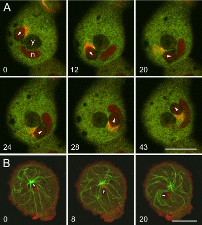
Actin polymerization can be induced in Dictyostelium by compressing the cells to bring phagosomes filled with large particles into contact with the plasma membrane. Asymmetric actin assembly results in rocketing movement of the phagosomes. We show that the compression-induced assembly of actin at the cytoplasmic face of the plasma membrane involves the Arp2/3 complex. We also identify two other proteins associated with the mechanically induced actin assembly. The class I myosin MyoB accumulates at the plasma membrane-phagosome interface early during the initiation of the response, and coronin is recruited as the actin filaments are disassembling. The forces generated by rocketing phagosomes are sufficient to push the entire microtubule apparatus forward and to dislocate the nucleus.


Figures

Figure 1.
Figure 1.
Phagosome rocketing in compressed cells viewed by TIRF microscopy to visualize protein recruitment to the cell cortex. Time in all figures is indicated in seconds after the initial frames. (A) Cell expressing LimEΔ-GFP to label filamentous actin structures (green), fed TRITC-labeled yeast particles (red), and compressed under an agarose sheet. When a phagosome contacted the cell cortex from the cytoplasmic face, actin assembled close to the plasma membrane. At one phagosome (indicated by an arrow), a ring of actin (20 s) became asymmetric as the phagosome moved first toward the bottom (72 and 98 s) and then toward the top of the images (138 and 178 s). At a second phagosome (open arrowhead), the site of actin deposition changed three times, and each time the direction of movement changed accordingly. These images are from Movie 1A; another example is shown in Movie 1B. (B) Cell expressing GFP-MyoB (green) and mRFP-LimEΔ (red), fed with unlabeled yeast (dark areas). When the agar overlay compressed the cell, this phagosome began a series of runs followed by pauses (Movie 2). Each time the phagosome paused, GFP-MyoB accumulated about the site of phagosome contact with the cortex (0 and 36 s, double arrows). Actin began assembling as a halo spread over a larger area of the cell cortex. The resumption of phagosome movement (6 and 53 s; the direction indicated by arrows) was associated with an asymmetric localization of MyoB and formation of an actin tail behind the transported particle. Bars, 5 μm.
Figure 2.
Figure 2.
Three-dimensional reconstruction of GFP-MyoB distribution (in green) relative to actin (mRFP-LimEΔ in red) at a phagosome arrested by pressure. The main panel shows an optical section in an X, Y plane close to the plasma membrane/phagosome interface. The left and upper panels represent cross-sections through the center of the phagosome in Z, Y and Z, X directions, respectively. Dual-color image stacks were recorded using a spinning disk confocal microscope with 0.3 μm Z-spacing. The rectangular scale bars indicate 2 μm in the Z direction and the same for the X, Y plane.
Figure 3.
Figure 3.
Localization of MyoB, Arp2/3, and F-actin at the plasma membrane in close contact with phagosomes as revealed by confocal microscopy. (A) A cell expressing GFP-MyoB and mRFP-LimEΔ. This cell has phagocytosed two yeast cells. It has also formed a macropinosome and is in the process of forming another (arrow). Note the similar relationships of MyoB and F-actin (LimEΔ) in the nascent macropinosome and in the areas surrounding the region of contact between the phagosomes and the plasma membrane. (B) Phagosomes in a cell expressing GFP-MyoB and mRFP-p41-Arc. At sites of phagosome–plasma membrane contact, the green ring of GFP-MyoB is surrounded by a red ring of Arp2/3. (C and D) The relative distribution of the Arp2/3 complex (GFP-Arp3) and F-actin (mRFP-LimEΔ) in two cells with rocketing phagosomes. There is almost complete overlap in the distribution of these two markers during phagosome movement, but with slightly greater enrichment of F-actin close to the phagosome membrane. As can be seen from the larger, clearer profiles of the yeast cells in C and D compared with A and B, the focal planes for C and D are slightly deeper in the cell. Movie 3 shows movement of the phagosome in C. The yeast cells used in these experiments were living cells of S. cerevisiae strain TH2–1B. Bars, 5 μm.
Figure 4.
Figure 4.
Persistence of the GFP-Arp3 label at the plasma membrane behind rocketing phagosomes revealed by confocal microscopy. For this experiment, the Dictyostelium clathrin light-chain mutant 2A1, which forms large cells, was used. Mutant cells expressing GFP-Arp3 were mixed with TRITC-labeled yeast and observed several hours later, after being covered with an agarose overlay. (A) On compressing the cells, rings of GFP-Arp3 appeared on the plasma membrane above and below the yeast-containing phagosomes. These rings were left behind as the phagosomes moved away. The images are from a confocal time series also shown in Movie 4. (B) In another cell, phagosome movement, initially back-and-forth, converted to a pinwheel trajectory. The last frame shows the pinwheeling phagosomes at another focal plane (Z) near the cell center. A field containing this cell is shown in Movie 5A; that time series continues with a higher magnification view of this cell in Movie 5B. Bars, 10 μm.
Figure 5.
Figure 5.
Sequential labeling of phagosome comet tails with LimEΔ and coronin viewed by confocal microscopy. A cell expressing mRFP-LimEΔ and coronin-GFP was fed living yeast ∼1 h before recording. The focal plane is close to the substratum, and the frames are separated by ∼4 s. (The same time series is shown in Movie 6.) Direction of movement is indicated in each frame by an arrowhead on the black area representing the yeast-containing phagosome. Each time the phagosome changed direction, mRFP-LimEΔ accumulated at its back. Between one frame and the next, the red patch became yellow or green, as coronin-GFP replaced the mRFP-LimEΔ labeling of actin filaments. (Compare frames 0 and 4 s; 8 and 12 s; and 24 and 28 s.) The yeast cells used in this experiment were living cells of S. cerevisiae strain TH2–1B. Bar, 5 μm.
Figure 6.
Figure 6.
Rocketing of phagosomes is separated in time from particle uptake and is not dependent on microtubules (confocal microscopy). (A) Rocketing phagosome in a cell expressing mRFP-LimEΔ and VatM-GFP, a subunit of the V-ATPase. The V-ATPase, which is delivered to the membrane of phagosomes a few minutes after uptake, is responsible for acidifying the endosomal lumen. The V-ATPase also populates membranes of the contractile vacuole complex. Close to the plasma membrane attached to the substratum, enrichment of mRFP-LimEΔ is evident at the rear of a rocketing phagosome (first two panels; arrowheads overlay the rocketing phagosome and show its direction of movement). The rocketing is also shown in Movie 7. In this focal plane, VatM-GFP is seen in the tubular network of the contractile vacuole system, which lies mostly on the plasma membrane. At the midplane of the cell (third panel, Z), the presence of VatM-GFP is evident in the phagosome membrane (arrows). No mRFP-LimEΔ labels the phagosome in this focal plane. The presence of VatM-GFP means that the phagosome is in the acidic (middle) stage of endocytic transit. (B) Rocketing in a nocodazole-treated cell expressing GFP-α-tubulin and mRFP-LimEΔ. The cells were fed yeast for 40 min and then incubated with 20 μM nocodazole. Two hours later, the cells were overlaid with agarose and observed. Phagosome movement still occurred, even though the microtubules had been depolymerized to short stubs. The yeast cells used in these experiments were living cells of S. cerevisiae strain TH2–1B. Bars, 10 μm.
Figure 7.
Figure 7.
Distortion of nucleus and microtubules by rocketing phagosomes. (A) Confocal view of a rocketing phagosome in a cell expressing GFP-MyoB and mRFP-LimEΔ. This phagosome made three circuits around a second larger phagosome (y) that was not moving and appears as a dark object near the cell center (Movie 8). One circuit is shown here. Each time the phagosome reached the last quadrant of the cell before nuclear impact, it accelerated briefly, momentarily outstripping its actin tail (panel 20). Subsequently, the phagosome rammed and displaced the nucleus (n), which appears slightly red because mRFP-LimEΔ has entered the nuclear matrix. Toward the end of Movie 8, the focus was shifted closer to the plasma membrane, revealing concentric rings of GFP-MyoB and mRFP-LimEΔ encircling the stationary phagosome. (B) Interactions between a rocketing phagosome and microtubules in a wild-type cell. AX2 cells expressing GFP-α-tubulin and mRFP-LimEΔ, were mixed with yeast about 2 h before the confocal microscopy time series shown in Movie 9 was captured. When the phagosome rammed the centrosome, it displaced the nucleus and stretched out the microtubules (panel 0), which relaxed when the phagosome turned away (panel 8). The microtubules were bent during lateral movements of the phagosome (panel 20). The taut appearance of the straightened microtubules suggests that they are anchored at the cell cortex as well as the centrosome. The yeast used for the experiment in Figure 7A were heat-killed, and those for the experiment in Figure 7B were living cells of S. cerevisiae strain 5288C, which are smaller than wild-type yeast cells. Bars, 10 μm.

Similar articles

Cited by

References

    1. Aubrey L., Klein G., Martiel J.-L., Satre M. Kinetics of endosomal pH evolution in Dictyostelium discoideum amoebae. Study by fluorescence spectroscopy. J. Cell Sci. 1993;105:861–866. - PubMed
    1. Axelrod D. Total internal reflection fluorescence microscopy in cell biology. Traffic. 2001;2:764–774. - PubMed
    1. Bernheim-Groswasser A., Wiesner S., Golsteyn R. M., Carlier M. F., Sykes C. The dynamics of actin-based motility depend on surface parameters. Nature. 2002;417:308–311. - PubMed
    1. Bretschneider T., Diez S., Anderson K., Heuser J., Clarke M., Müller-Taubenberger A., Köhler J., Gerisch G. Dynamic actin patterns and Arp2/3 assembly at the substrate-attached surface of motile cells. Curr. Biol. 2004;14:1–10. - PubMed
    1. Cai L., Holoweckyj N., Schaller M. D., Bear J. E. Phosphorylation of coronin 1B by protein kinase C regulates interaction with Arp2/3 and cell motility. J. Biol. Chem. 2005;280:31913–31923. - PubMed

Publication types

LinkOut - more resources