. 2006 Sep 13;26(37):9413-25.
doi: 10.1523/JNEUROSCI.2591-06.2006.

Receptor and transmitter release properties set the time course of retinal inhibition

Affiliations

Receptor and transmitter release properties set the time course of retinal inhibition

Erika D Eggers et al. J Neurosci. .

Abstract

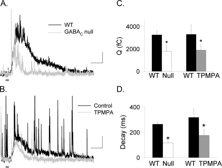
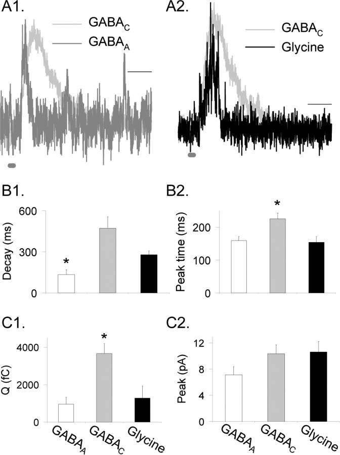
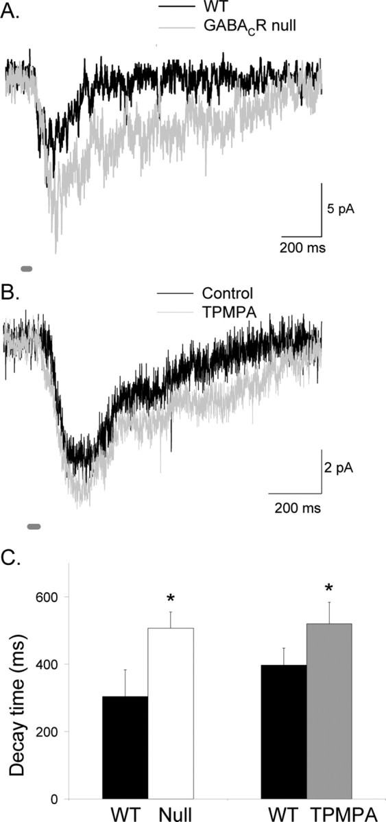
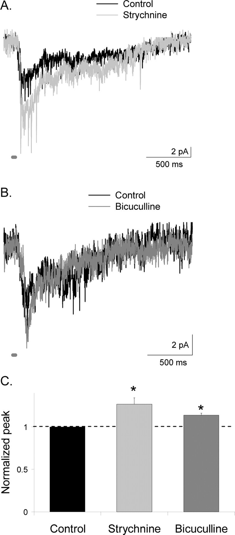
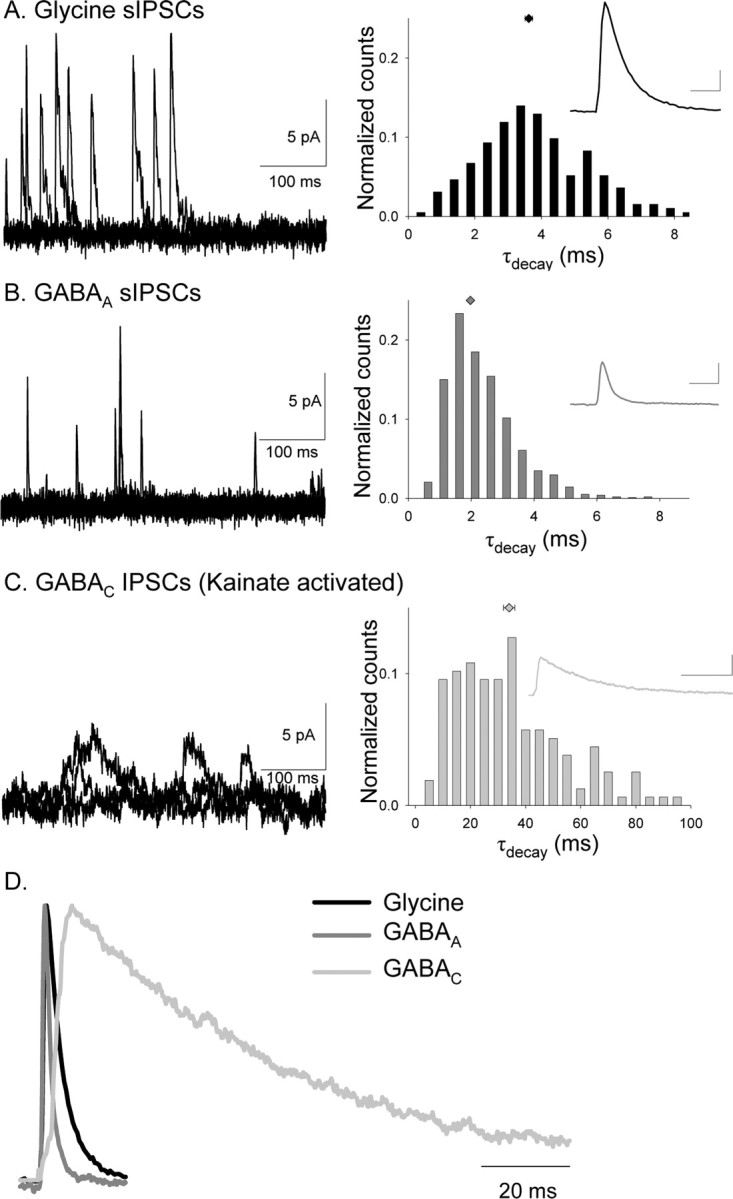
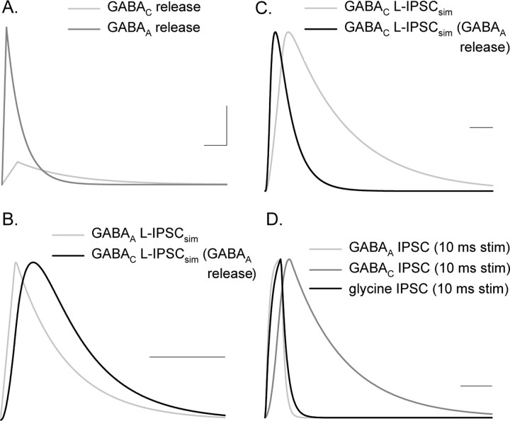
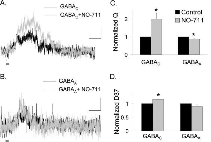
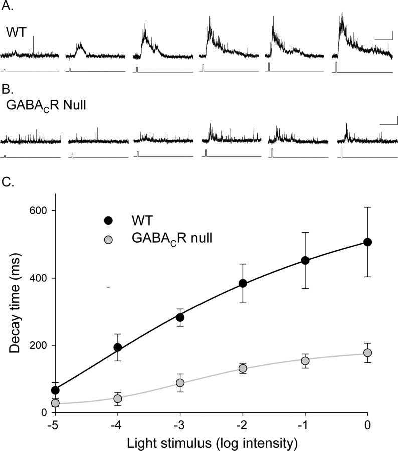
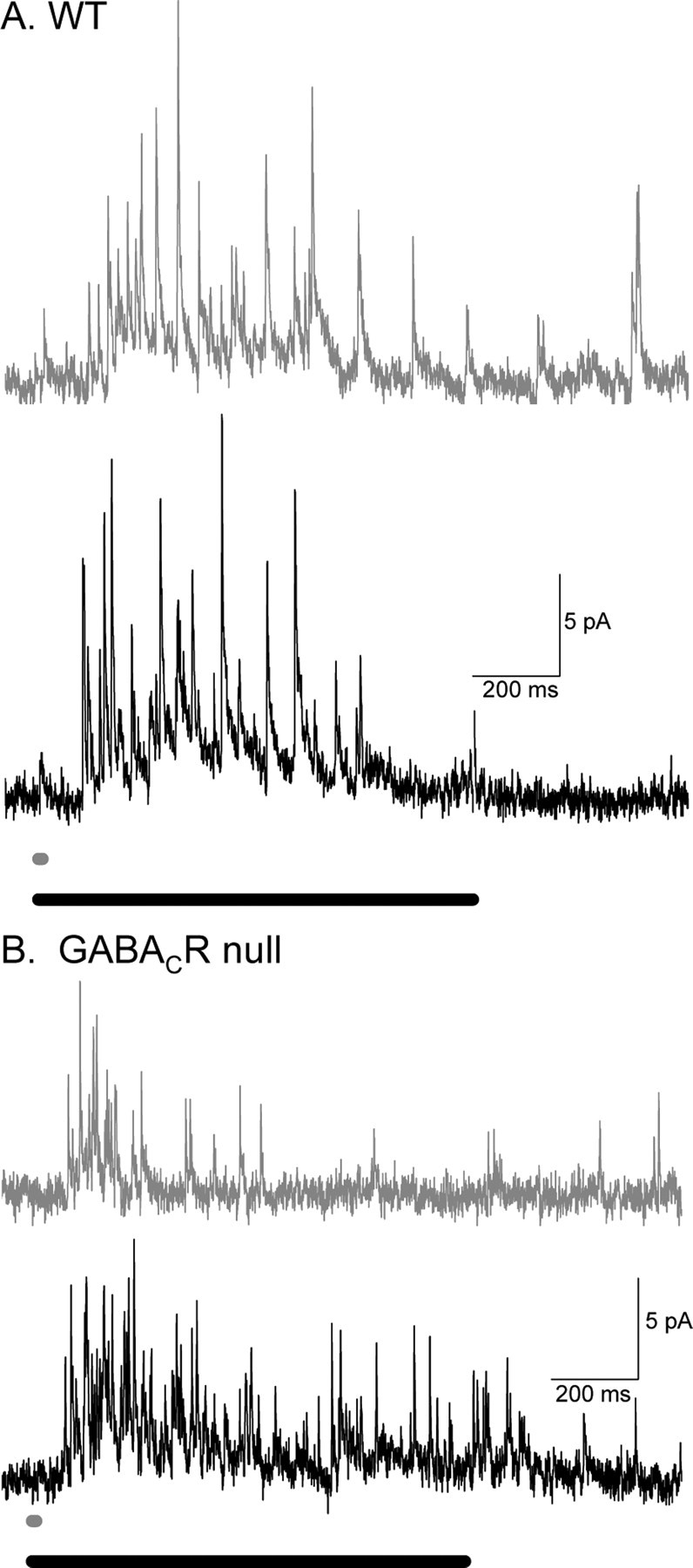
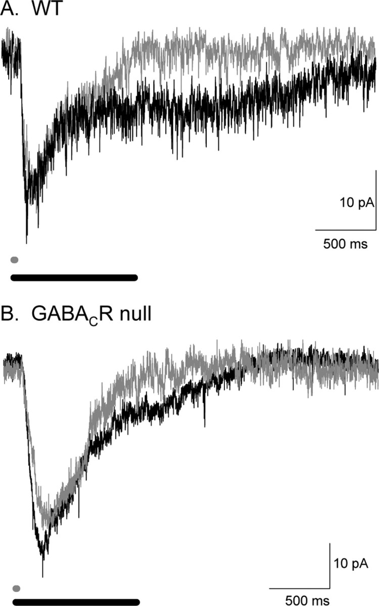
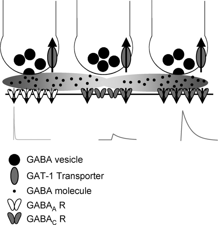
Synaptic inhibition is determined by the properties of postsynaptic receptors, neurotransmitter release, and clearance, but little is known about how these factors shape sensation-evoked inhibition. The retina is an ideal system to investigate inhibition because it can be activated physiologically with light, and separate inhibitory pathways can be assayed by recording from rod bipolar cells that possess distinct glycine, GABA(A), and GABA(C) receptors (R). We show that receptor properties differentially shape spontaneous IPSCs, whereas both transmitter release and receptor properties shape light-evoked (L) IPSCs. GABA(C)R-mediated IPSCs decayed the slowest, whereas glycineR- and GABA(A)R-mediated IPSCs decayed more rapidly. Slow GABA(C)Rs determined the L-IPSC decay, whereas GABA(A)Rs and glycineRs, which mediated rapid onset responses, determined the start of the L-IPSC. Both fast and slow inhibitory inputs distinctly shaped the output of rod bipolar cells. The slow GABA(C)Rs truncated glutamate release, making the A17 amacrine cell L-EPSCs more transient, whereas the fast GABA(A)R and glycineRs reduced the initial phase of glutamate release, limiting the peak amplitude of the L-EPSC. Estimates of transmitter release time courses suggested that glycine release was more prolonged than GABA release. The time course of GABA release activating GABA(C)Rs was slower than that activating GABA(A)Rs, consistent with spillover activation of GABA(C)Rs. Thus, both postsynaptic receptor and transmitter release properties shape light-evoked inhibition in retina.


Figures

Figure 1.
Figure 1.
GABAC receptors prolong rod bipolar cell L-IPSCs. A, L-IPSCs evoked by brief light stimuli (30 ms; thick dark gray bar) were smaller and decayed faster in GABACR null mice (gray trace) than in WT mice (black trace). Calibration: 5 pA, 200 ms. B, Blocking GABAC receptors with TPMPA decreased and shortened L-IPSCs (gray trace) compared with control conditions (black trace). C, The Q in GABACR null mice was significantly smaller than in WT mice (*p < 0.05). The Q in rod bipolar cells after TPMPA application was significantly smaller than in control retinas (*p < 0.05). D, The L-IPSC decay (D37) in GABACR mice was significantly briefer than for WT mice (*p < 0.001). Similarly, the L-IPSC decay measured in the presence of TPMPA also was briefer than that measured in control conditions (*p < 0.05).
Figure 2.
Figure 2.
Glycine, GABAA, and GABAC receptors mediated L-IPSCs, with different properties. A1 and A2 show pharmacologically isolated and peak-scaled L-IPSCs evoked by a 30 ms light stimulus (dark gray bar at bottom of trace) mediated by GABAA, GABAC, and glycine receptors. GABAC receptors mediated L-IPSCs with a slower decay time than those mediated by GABAA or glycine receptors. Calibration, 200 ms. B1, The D37 was measured from L-IPSCs mediated by isolated glycine (black), GABAA (white), and GABAC (gray) receptors. GABAA L-IPSCs had a significantly briefer D37 than either glycine receptors or GABAC receptors; *p < 0.01. Glycine receptor-mediated L-IPSCs were on average briefer than GABAC receptor-mediated L-IPSCs (p < 0.08). B2, GABAC receptor-mediated L-IPSCs had a longer time-to-peak than either glycine or GABAA receptor-mediated L-IPSCs; *p < 0.01. There was no significant difference in time-to-peak between GABAA and glycine L-IPSCs (p = 0.8). C1, GABAC receptor-mediated L-IPSCs had significantly larger Q than either GABAA or glycine L-IPSCs (p < 0.01). There was no significant difference in Q between GABAA and glycine L-IPSCs (p = 0.3). C2, GABAA receptor-mediated L-IPSCs on average had a smaller peak value than GABAC and glycine L-IPSCs (p < 0.1).
Figure 3.
Figure 3.
Presynaptic GABAC receptors make L-EPSCs from A17 amacrine cells more transient. A, Shown are L-EPSCs (30 ms light stimulus; dark gray bay) from A17 amacrine cells from WT and GABACR null mice. The absence of GABAC receptors in GABACR null mice causes the L-EPSC to have a longer decay and larger charge transfer. B, A similar effect was observed in WT mice when TPMPA was added to block GABAC receptors. C, The decay (D37) of A17 amacrine cells from GABACR null mice (*p < 0.05) and WT mice in TPMPA (*p < 0.01) was significantly longer than WT mice in control conditions.
Figure 4.
Figure 4.
Presynaptic glycine and GABAA receptors decrease the peak response of L-EPSCs from A17 amacrine cells. A, The peak amplitude of L-EPSCs (30 ms light stimulus; dark gray bar) from A17 amacrine cells from GABACR null mice was increased by the addition of strychnine to block glycine receptors, but the decay time of the response was unaffected. B, Similarly, the peak amplitude of L-EPSCs from A17 amacrine cells from GABACR null mice was increased by addition of bicuculline to block GABAA receptors, but the response decay was unaffected. C, The amplitude of L-EPSCs was significantly larger with the addition of both strychnine (*p < 0.05) and bicuculline (*p < 0.05). Peak values in bicuculline and strychnine are normalized to control values, represented by the dotted line.
Figure 5.
Figure 5.
Glycine, GABAA, and GABAC receptors mediate sIPSCs with distinct kinetics in rod bipolar cells. A, Examples of isolated glycine receptor sIPSCs that were measured in the presence of bicuculline and TPMPA (left). The τdecay histogram distribution (normalized to the total number of events) for all glycine receptor-mediated sIPSCs recorded are shown in the right, and the inset shows the average glycine receptor-mediated sIPSC. Calibration:2 pA, 5 ms. B, Examples of isolated GABAA receptor sIPSCs that were measured in the presence of strychnine and TPMPA (left). The normalized τdecay histogram distribution for all GABAA receptor-mediated sIPSCs recorded are shown in the right, and the inset shows the average GABAA receptor-mediated sIPSC. Calibration: 2 pA, 5 ms. GABAA receptor-mediated sIPSCs had a significantly shorter τdecay than glycine receptor-mediated sIPSCs (K–S, p < 0.0001). C, Examples of isolated GABAC receptor sIPSCs, measured in the presence of kainate, strychnine, and bicuculline (left). The normalized τdecay histogram distribution for all GABAC receptor-mediated sIPSCs recorded is shown in the right, and the inset shows the average GABAC receptor-mediated sIPSC. Calibration: 2 pA, 50 ms. GABAC receptor-mediated sIPSCs had a significantly longer τdecay than glycine (K–S, p < 0.0001) and GABAA (K–S, p < 0.0001) receptor-mediated sIPSCs. D, Average normalized sIPSCs mediated by glycine, GABAA, and GABAC receptors. GABAC receptor-mediated sIPSCs have the longest decay time.
Figure 6.
Figure 6.
GABAA and GABAC receptor-mediated L-IPSCs have distinct apparent release functions and receptor kinetics, both of which contribute to L-IPSC kinetics. A, Release functions computed by deconvolving idealized GABAA and GABAC receptor-mediated L-IPSCs (supplemental data, available at www.jneurosci.org as supplemental material). GABAA receptor-mediated L-IPSCs have a much larger release function than GABAC receptors, likely because of the much smaller Q of GABAA sIPSCs versus GABAC sIPSCs. The GABAC release function has a prolonged tail not shown by the GABAA release function. Calibration: 0.05 quanta/ms, 200 ms. B, To determine how much of the difference between GABAA and GABAC L-IPSC kinetics was attributable to receptor properties, we used the GABAA calculated release function to simulate L-IPSCs with the GABAA and GABAC sIPSCs. Shown is the normalized GABAC L-IPSC, using the GABAA release function, that had a slower rise and decay time than the normalized GABAA L-IPSC, as a result of the slower kinetics of the GABAC sIPSCs. The L-IPSC simulated with the GABAA release function and sIPSC matched the properties of the average GABAA L-IPSC from Table 1. Calibration, 200 ms. C, Shown are the normalized GABAC L-IPSCs using the calculated GABAA and GABAC release rates. The prolonged tail of the estimated GABAC release rate slows the kinetics of the GABAC L-IPSCs. Again, the L-IPSC simulated with the GABAC release function and sIPSC matched the properties of the average GABAC L-IPSC from Table 1. Calibration, 200 ms. D, If GABAA, GABAC, and glycine receptor sIPSCs are activated by a brief (square wave, lasting 10 ms) burst of neurotransmitter, then the distinct receptor properties filter the response, as shown by these normalized responses. Calibration, 200 ms.
Figure 7.
Figure 7.
GABAC receptor-mediated L-IPSCs are enhanced by blocking GABA uptake, but GABAA receptor-mediated L-IPSCs are not. A, GABAC receptor-mediated L-IPSCs (30 ms light) are enhanced by NO-711 (5 μm), which inhibits the GAT-1 GABA transporter. Calibration: 2 pA, 200 ms. B, GABAA receptor-mediated L-IPSCs (30 ms light) are minimally changed by the addition of NO-711. C, GABAC L-IPSCs Q is significantly increased by NO-711 (*p < 0.05), whereas the GABAA L-IPSC Q is decreased by a small amount (*p < 0.01). D, The D37 of GABAC receptor-mediated L-IPSCs was increased by NO-711 (*p < 0.01), whereas the D37 of GABAA receptor-mediated L-IPSCs was unchanged (p = 0.3).
Figure 8.
Figure 8.
Eliminating GABAC receptors reduced the decay time of rod bipolar cell L-IPSCs as a function of light intensity. A, L-IPSCs from a WT rod bipolar cell in response to increasing intensities of light (30 ms light stimulus). Calibration: 5 pA, 500 ms. B, L-IPSCs from a GABACR null rod bipolar cell in response to increasing intensities of light. The increases in the magnitude and decay of the response are smaller for the GABACR null mice. C, The average L-IPSC D37 values from WT (n = 10) and GABACR null (n = 6) mice are plotted as a function of light intensity (log relative intensity). The curves are the best fits to a logistic function. The maximum D37 of the GABACR null curve was 35% of the WT maximum. The L20–80 values of the fits were not significantly changed (WT, 2.1 ± 0.8; null, 2.4 ± 0.8), but the GABACR null curve had a lower EC50 (WT, −3.4 ± 0.2; null, −2.7 ± 0.2).
Figure 9.
Figure 9.
WT and GABACR null mice L-IPSCs respond differently to increasing light duration. A, Example L-IPSCs from WT rod bipolar cells in response to 10 ms (dark gray trace) and 1000 ms (black trace) light stimulation. The two L-IPSCs do not have drastically different charge transfers. B, Example L-IPSCs from GABACR null rod bipolar cells in response to 10 ms (dark gray trace) and 1000 ms (black trace) light stimulation. The 10 ms L-IPSC is much smaller than that 1000 ms L-IPSC. GABACR null L-IPSCs increase significantly more when the light duration is increased (p < 0.01).
Figure 10.
Figure 10.
L-IPSCs differentially shape A17 L-EPSCs in WT and GABACR null mice. A, Example L-EPSCs from WT A17 amacrine cells in response to 10 ms (dark gray traces) and 1000 ms (black traces) light stimulation. The 10 ms L-EPSC is smaller than that 1000 ms L-EPSC. B, Example L-EPSCs from GABACR null A17 amacrine cells in response to 10 and 1000 ms light stimulation. The two L-EPSCs do not have as large a difference in charge transfers as the WT L-EPSCs. WT L-EPSCs increase significantly more when the light duration is increased (p < 0.01).
Figure 11.
Figure 11.
The properties of inhibitory synaptic transmission are controlled by many independent factors. Shown is an example of three amacrine cell terminals releasing GABA onto synapses on a bipolar cell. The terminal on the left is activating GABAA receptors that have a short time course, and the terminal on the right is activating GABAC receptors that have a prolonged time course. The terminal in the middle has not released GABA, but the GABAC receptors underlying it are being activated by spillover from neighboring synapses, which respond with a small, delayed current. GABA is taken up by GAT-1 transporters, which control the amount of GABA that leaves the synapse.

Similar articles

Cited by

References

    1. Amin J, Weiss DS. Homomeric rho 1 GABA channels: activation properties and domains. Receptors Channels. 1994;2:227–236. - PubMed
    1. Barbour B. An evaluation of synapse independence. J Neurosci. 2001;21:7969–7984. - PMC - PubMed
    1. Berntson A, Smith RG, Taylor WR. Postsynaptic calcium feedback between rods and rod bipolar cells in the mouse retina. Vis Neurosci. 2004;21:913–924. - PubMed
    1. Bloomfield SA, Xin D. Surround inhibition of mammalian AII amacrine cells is generated in the proximal retina. J Physiol (Lond) 523. 2000;3:771–783. - PMC - PubMed
    1. Brickley SG, Cull-Candy SG, Farrant M. Development of a tonic form of synaptic inhibition in rat cerebellar granule cells resulting from persistent activation of GABAA receptors. J Physiol (Lond) 1996;497:753–759. - PMC - PubMed

Publication types

MeSH terms

LinkOut - more resources