. 2006 Sep 13;26(37):9503-11.
doi: 10.1523/JNEUROSCI.2021-06.2006.

Context-dependent human extinction memory is mediated by a ventromedial prefrontal and hippocampal network

Affiliations

Context-dependent human extinction memory is mediated by a ventromedial prefrontal and hippocampal network

Raffael Kalisch et al. J Neurosci. .

Abstract

In fear extinction, an animal learns that a conditioned stimulus (CS) no longer predicts a noxious stimulus [unconditioned stimulus (UCS)] to which it had previously been associated, leading to inhibition of the conditioned response (CR). Extinction creates a new CS-noUCS memory trace, competing with the initial fear (CS-UCS) memory. Recall of extinction memory and, hence, CR inhibition at later CS encounters is facilitated by contextual stimuli present during extinction training. In line with theoretical predictions derived from animal studies, we show that, after extinction, a CS-evoked engagement of human ventromedial prefrontal cortex (VMPFC) and hippocampus is context dependent, being expressed in an extinction, but not a conditioning, context. Likewise, a positive correlation between VMPFC and hippocampal activity is extinction context dependent. Thus, a VMPFC-hippocampal network provides for context-dependent recall of human extinction memory, consistent with a view that hippocampus confers context dependence on VMPFC.


Figures

Figure 1.
Figure 1.
AB–AB design. On day 1, subjects were fear conditioned to a CS+ (a face) through multiple pairings with a UCS (electric shock) in context A (conditioning context, block A1). Fear responses were extinguished in context B (extinction context, block B1) through multiple CS+ presentations in the absence of the UCS. This procedure was repeated in additional blocks (blocks A2, B2), leading to robust context discrimination. As a control for nonassociative effects, we also used a nonpredictive CS− (a face of opposite gender) that was never paired with the UCS and presented intermixed with the CS+. Contexts were defined by screen color (black in one context, changing between red and orange in the other) and auditory input (two sounds, changing synchronously with the color of the screen in the red–orange context only). These marked physical differences between the two contexts were intended to facilitate context discrimination. On day 2, subjects were tested for CS+-induced recall of fear and extinction memories in both contexts, each context being presented 16 times in alternating order. Recall of fear memory in context A on day 2 was facilitated by additionally presenting one unpaired shock at the beginning of each context A block, thus again firmly associating context A with the UCS. The task was a speeded gender decision task in response to the face stimuli. Gender of faces, conditioning, and extinction contexts and the order of those contexts on day 2 were counterbalanced across subjects. Lightning bolt, Electric shock.
Figure 2.
Figure 2.
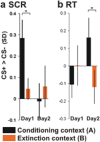
Successful discrimination learning and context-dependent recall. a, SCR. Larger SCRs to the CS+ versus CS− during fear conditioning in context A compared with the extinction context B on day 1 indicate learning of the CS+–UCS contingency (fear memory) in context A and of the CS+–noUCS contingency (extinction memory) in context B on day 1 (left). b, RTs. Slower RTs to the CS+ versus CS− in the conditioning context A compared with the extinction context B on day 2 indicate recall of fear memory in context A and recall of extinction memory in B on day 2 (right). Scale: z scores (unit, SDs). z scores were used instead of raw values (micro-ohms, seconds) to account for interindividual differences. *p < 0.05, one-tailed.
Figure 3.
Figure 3.
Recall of fear memory. Activations associated with recall of fear memory on day 2. a, Right (R) posterior hippocampus [MNI coordinates (38, −32, −12)]. b, Left (L) amygdala [coordinates (−28, −2, −32)]. Images show group-level estimates for the contrast (CS+ > CS−)A, modulated by RT, display threshold p ≤ 0.01. Activations superimposed on mean structural image, masked for hippocampus and amygdala. Insets, Group estimates for CS+ > CS− contrasts in both contexts.
Figure 4.
Figure 4.
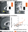
Recall of extinction memory. a, Activations associated with context-dependent recall of extinction memory on day 2. Images show contrast (CS+ > CS−)B > (CS+ > CS−)A, display threshold p ≤ 0.001, masked for VMPFC and hippocampus. Activations are superimposed on mean structural image. Insets, Group-level estimates for CS+ > CS− contrasts in both contexts. b, CS+-evoked left VMPFC activity is positively correlated with CS+-evoked left anterior hippocampus activity in the extinction context only. Left, Correlation in VMPFC in the extinction context, display threshold p ≤ 0.001, masked by VMPFC activation in a. Right, An extinction context-specific correlation is apparent from the subject-specific estimates for the two CS+ > CS− contrasts (black: conditioning context, r = 0.212, p = 0.207, one-tailed; orange: extinction context, r = 0.715, p = 0.0005, one-tailed). L, Left.

Similar articles

Cited by

References

    1. Amaral DG. Introduction: what is where in the medial temporal lobe? Hippocampus. 1999;9:1–6. - PubMed
    1. Anagnostaras SG, Gale G, Fanselow MS. Hippocampus and contextual fear conditioning: recent controversies and advances. Hippocampus. 2001;11:8–17. - PubMed
    1. Ashburner J, Friston KJ, Penny W. Imaging neuroscience—theory and analysis. In: Frackowiak RS, Friston KJ, Frith C, Dolan RJ, Price CJ, editors. Human brain function. San Diego: Academic; 2004. pp. 599–1104.
    1. Blair HT, Huynh VK, Vaz VT, Van J, Patel RR, Hiteshi AK, Lee JE, Tarpley JW. Unilateral storage of fear memories by the amygdala. J Neurosci. 2005;25:4198–4205. - PMC - PubMed
    1. Boshuisen ML, Ter Horst GJ, Paans AM, Reinders AA, Den Boer JA. rCBF differences between panic disorder patients and control subjects during anticipatory anxiety and rest. Biol Psychiatry. 2002;52:126–135. - PubMed

Publication types

MeSH terms