

Effects of number of diffusion gradient directions on derived diffusion tensor imaging indices in human brain
- PMID: 16971635
- PMCID: PMC8139764
Effects of number of diffusion gradient directions on derived diffusion tensor imaging indices in human brain
Abstract
Background and purpose: The effects of a number of diffusion-encoding gradient directions (NDGD) on diffusion tensor imaging (DTI) indices have been studied previously with theoretic analysis and numeric simulations. In this study, we made in vivo measurements in the human brain to compare different clinical scan protocols and to evaluate their effects on the calculated DTI indices.
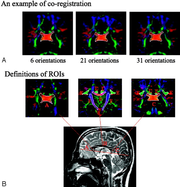
Methods: Fifteen healthy volunteers were scanned with a 1.5T MR scanner. Single-shot DTI images were acquired using 3 protocols different in NDGD and number of excitations (NEX) for each direction (NDGD/NEX = 6/10, 21/3, 31/2). Means and standard error of mean (SEM) were calculated and compared in 6 regions of interest (ROIs) for mean diffusivity (D), fractional anisotropy (FA), diffusion tensor eigenvalues (lambda(1), lambda(2), and lambda(3)), and correlation coefficients (r) of these indices among the 3 DTI protocols.
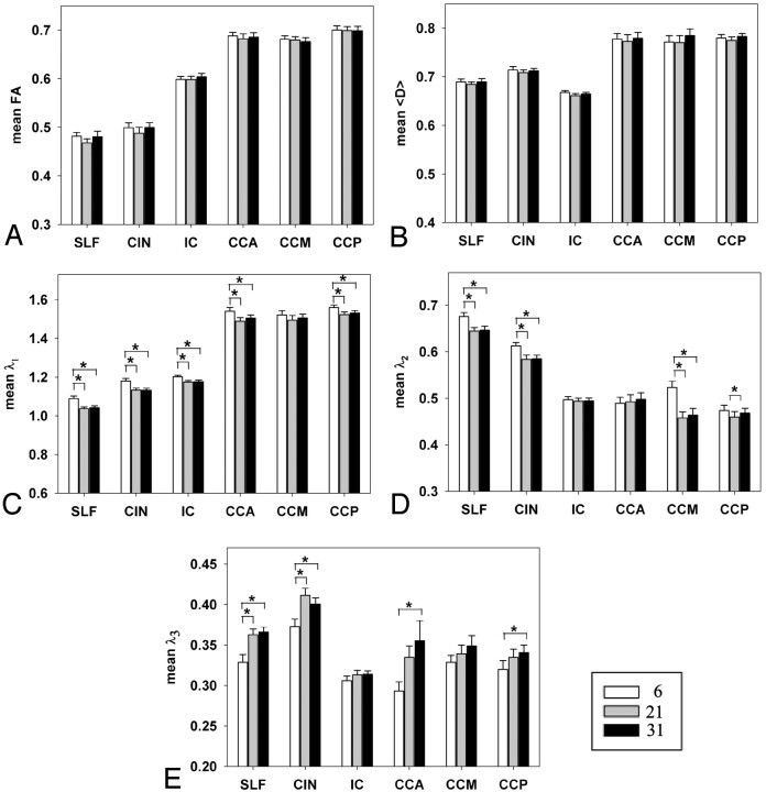
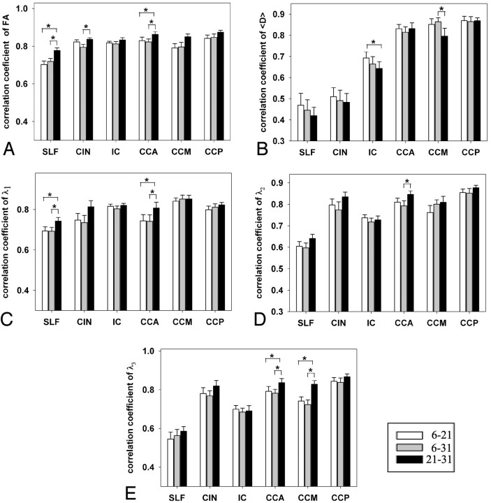
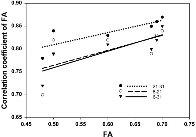
Results: At the ROI level, no significant differences were found for the mean and SEM of D and FA among protocols (P > .05). The 6-NDGD protocol, however, yielded higher values for lambda(1) and lambda(2) and lower values for lambda(3) in most ROIs (P < .05) compared with the other protocols. At the voxel level, the correlation between the protocols r(21-31) were higher than r(6-21) and r(6-31) in most ROIs. The correlation of FA among 3 protocols also increased with increasing anisotropy.
Conclusion: For ROI analyses, different NDGDs lead to similar values of FA and D but different eigenvalues. However, different NDGDs at the voxel level provide varying values. The selection of the NDGD, therefore, should depend on the focus of different DTI applications.
Figures
References
-
- Takahashi S, Yonezawa H, Takahashi J, et al. Selective reduction of diffusion anisotropy in white matter of Alzheimer disease brains measured by 3.0 Tesla magnetic resonance imaging. Neurosci Lett 2002;332:45–48 - PubMed
-
- Fellgiebel A, Wille P, Muller MJ, et al. Ultrastructural hippocampal and white matter alterations in mild cognitive impairment: a diffusion tensor imaging study. Dement Geriatr Cogn Disord 2004;18:101–08 - PubMed
-
- Zhong J, Ni H, Zhu T, et al. MR diffusion tensor imaging (DTI) and neuropsychological testing for neuronal connectivity in Alzheimer’s disease (AD) patients. Proceedings of SPIE: Medical Imaging 2004—Physiology, Function, and Structure from Medical Images 2004;5369:238–49
-
- Coombs BD, Best A, Brown MS, et al. Multiple sclerosis pathology in the normal and abnormal appearing white matter of the corpus callosum by diffusion tensor imaging. Mult Scler 2004;10:392–97 - PubMed
Publication types
MeSH terms
Grants and funding
LinkOut - more resources
Full Text Sources