. 2006 Nov;44(11):4163-71.
doi: 10.1128/JCM.01137-06. Epub 2006 Sep 13.

Molecular characterization of disease-associated streptococci of the mitis group that are optochin susceptible

Affiliations

Molecular characterization of disease-associated streptococci of the mitis group that are optochin susceptible

Luz Balsalobre et al. J Clin Microbiol. 2006 Nov.

Abstract

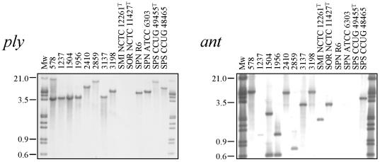
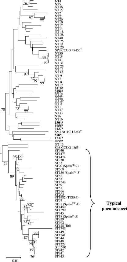
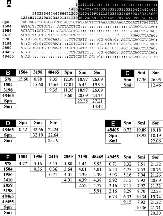
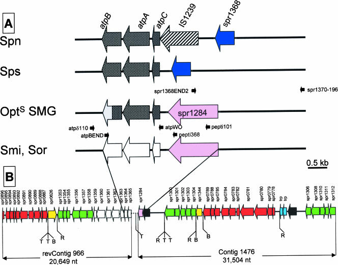
Eight optochin-susceptible (Opt(s)) alpha-hemolytic (viridans) streptococcus isolates were characterized at the molecular level. These isolates showed phenotypic characteristics typical of both viridans streptococci and Streptococcus pneumoniae. Comparison of the sequence of housekeeping genes from these isolates with those of S. pneumoniae, Streptococcus mitis, Streptococcus oralis, and Streptococcus pseudopneumoniae suggested that the Opt(s) isolates corresponded to streptococci of the mitis group. Besides, the Opt(s) streptococci were negative by a Gen-Probe AccuProbe pneumococcus test and hybridized with specific pneumococcal probes (lytA and ply) but also with ant, a gene not present in most S. pneumoniae strains. Moreover, the isolates were insoluble in 1% sodium deoxycholate but completely dissolved in 0.1% deoxycholate. Sequence analysis of the lytA gene revealed that the Opt(s) streptococci carried lytA alleles characteristic of those present in nonpneumococcal streptococci of the mitis group. The determination of the partial nucleotide sequence embracing the atp operon encoding the F(o)F(1) H(+)-ATPase indicated that the optochin susceptibility of the isolates was due to the acquisition of atpC, atpA, and part of atpB from S. pneumoniae by horizontal gene transfer.


Figures

FIG. 1.
FIG. 1.
Southern blot hybridization of streptococcal isolates by hybridization with ply- and ant-specific probes. Chromosomal DNAs were cleaved with ClaI (A) and EcoRV plus NcoI (B), and the fragments were separated in 1% agarose gels. The gel was blotted, and the blot was probed with biotinylated DNA as follows: an insert of plasmid pJCP191 containing the ply gene and a PCR fragment from S. pneumoniae 3870 containing most of the ant gene. Far-left and far-right lanes, biotinylated DNA ladder. Numbers at left are molecular size markers in kilodaltons. SMI, S. mitis; SOR, S. oralis; SPN, S. pneumoniae; SPS, S. pseudopneumoniae.
FIG. 2.
FIG. 2.
Dendrogram of genetic relationships between the SMG isolates examined in this study (shown in boldface type and labeled with an asterisk) and other streptococcal isolates. The dendrogram was constructed from housekeeping gene sequence data by using the neighbor-joining method. Only bootstrap confidence values of ≥70% are shown. NT and ST denote nontypeable SMG and sequence type, respectively (11). The scale represents the number of nucleotide substitutions per site. SPS, S. pseudopneumoniae; SMI, S. mitis.
FIG. 3.
FIG. 3.
Polymorphism in the atpC, atpA, and atpB genes of Opts SMG. (A) The nucleotides present at each polymorphic site are shown for S. pneumoniae R6, but for the other strains, only nucleotides that differ from those in R6 are shown. Nucleotide positions at the 1,499-bp fragments are indicated vertically above the sequences. Nucleotide positions located upstream of atpC, in atpA, or in atpB are indicated on a white, gray, or black background, respectively. Colons indicate nucleotides identical to those of strain R6. SMG strains are identified by numbering corresponding to that in Table 1. (B to F) Pairwise comparison of the nucleotide sequences located upstream of atpC (nucleotide positions 1 to 114) (B), in atpC (nucleotide positions 115 to 315) (C), in atpA (nucleotide positions 350 to 1066) (D), in the conserved part of atpB (nucleotide positions 1080 to 1217) (E), and in the most divergent region of atpB (nucleotide positions 1218 to 1499) (F). Matrices of PEDs between aligned sequences are shown. Abbreviations: Spn, S. pneumoniae R6; Smi, type strain of S. mitis; Sor, type strain of S. oralis.
FIG. 4.
FIG. 4.
Genetic structure of the atp region and its surrounding regions in SMG. Big arrows indicate the genes and their direction of transcription, using as a reference the S. pneumoniae genome (17, 37). The oligonucleotides used in PCR experiments are indicated by small black arrows. (A) Crosshatched and open arrows correspond to the atp genes of S. pneumoniae (Spn)/S. pseudopneumoniae (Sps) or S. mitis (Smi)/S. oralis (Sor), respectively. The atpB gene from Opts SMG is represented as a doubly shaded arrow, where the gray color indicates sequence divergence from either S. pneumoniae or S. mitis genes. (B) Partial genomic map of the DNA regions flanking the S. mitis type strain operon. The DNA region linking contig 966, whose complementary and inverted sequence is represented (revContig 966), and contig 1476 is represented as a dotted line; it has been sequenced previously (23), and it corresponds to the S. mitis/S. oralis scheme shown in panel A. Genes are named according to their S. pneumoniae R6 homologues. Yellow and pink arrows indicate genes that are translocated compared with their location and orientation in S. pneumoniae. Red arrows indicate gene or gene clusters that are inverted. S. mitis genes showing synteny with those of S. pneumoniae are indicated by green or open (for the atp genes) arrows. Light-blue and filled arrows represent transposase genes and genes lacking any significant similarity with those included in the databases, respectively. T, B, and R indicate the locations of transposase genes/IS-like elements and BOX or RUP repeats, respectively, as annotated in the S. pneumoniae R6 genome sequence (17). nt, nucleotides.

Similar articles

Cited by

References

    1. Abrahams, J. P., A. G. W. Leslie, and J. E. Walker. 1994. Structure at 2.8 Å resolution of F1-ATPase from bovine heart mitochondria. Nature 370:621-628. - PubMed
    1. Altschul, S. F., T. L. Madden, A. A. Schaffer, J. Zhang, Z. Zhang, W. Miller, and D. J. Lipman. 1997. Gapped BLAST and PSI-BLAST: a new generation of protein database search programs. Nucleic Acids Res. 25:3389-3402. - PMC - PubMed
    1. Arbique, J. C., C. Poyart, P. Trieu-Cuot, G. Quesne, M. da Glória S. Carvalho, A. G. Steigerwalt, R. E. Morey, D. Jackson, R. J. Davidson, and R. R. Facklam. 2004. Accuracy of phenotypic and genotypic testing for identification of Streptococcus pneumoniae and description of Streptococcus pseudopneumoniae sp. nov. J. Clin. Microbiol. 42:4686-4696. - PMC - PubMed
    1. Balsalobre, L., M. J. Ferrándiz, J. Liñares, F. Tubau, and A. G. de la Campa. 2003. Viridans group streptococci are donors in horizontal transfer of topoisomerase IV genes to Streptococcus pneumoniae. Antimicrob. Agents Chemother. 47:2072-2081. - PMC - PubMed
    1. Borek, A. P., D. C. Dressel, J. Hussong, and L. R. Peterson. 1997. Evolving clinical problems with Streptococcus pneumoniae: increasing resistance to antimicrobial agents, and failure of traditional optochin identification in Chicago, Illinois, between 1993 and 1996. Diagn. Microbiol. Infect. Dis. 29:209-214. - PubMed

Publication types

LinkOut - more resources