

doi: 10.1901/jaba.2005.1-27.
Molecular Basis of Steroid Action in the Prostate
Affiliations
- PMID: 16971966
- PMCID: PMC1564164
- DOI: 10.1901/jaba.2005.1-27
Item in Clipboard
Molecular Basis of Steroid Action in the Prostate
Cellscience.
.
No abstract available
Figures
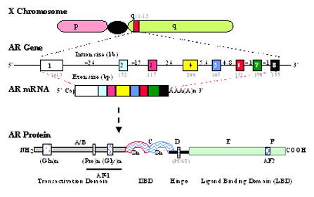
Schematic illustration of the location, exon structure and protein domain structure of the AR gene. (Top) The location of AR gene at the q11-12 of X chromosome. (Middle) The AR gene and its mRNA. The AR gene consists of 8 exons (boxes) and 7 introns (line), and the size of each exon and introns is indicated in kbases. (Bottom) The AR protein. The domains of AR are indicated. Relative positions of glutamine (Gln), proline (Pro) and glycine (Gly) repeats within the N-terminal domain are shown by the indicated boxes. The transactivation function domains, AF-1 and AF-2 are located within the N-terminal domain and ligand-binding domain, respectively. Two zinc fingers in the DNA binding domain and a PEST sequence in hinge region are indicated.
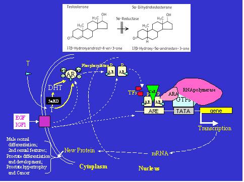
Illustrations of the molecular events of androgen-AR action in a target cell, and the 5α-reductase action. When testosterone (T) enters the cell, it can be converted to dihydrotestosterone (DHT) in the cell by 5α-reductases (5αRD) (top panel). Both T and DHT bind to androgen receptor (AR), resulting in a conformational change in AR and translocation of the receptor complex to nucleus. This complex interacts with androgen response element (ARE) on the target gene, and regulates gene expression in concert with co-regulators (CoR), transcription factors (TF) and the general transcription complex. The changes in androgen-target proteins in the cell eventually affect cellular structure and function related to male sexual differentiation, physiology, and pathophysiology. The function of AR can be activated or modified by non-ligand factors such as growth factors, EGF and IGF-1. GTFs – general transcription factors; ARA – androgen receptor associated proteins; P- phosphorylation; TFs – transcription factors; hsp – heat shock protein.
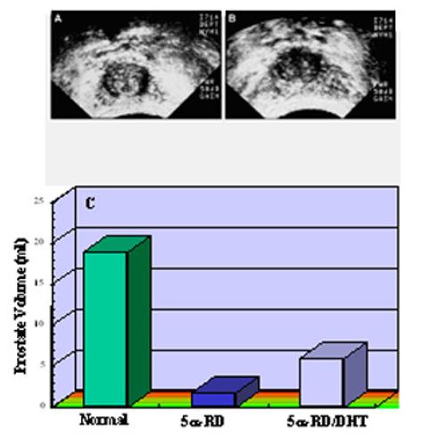
Comparison of prostate sizes between age-matched normal adult males and 5α-reductase-2 deficient patients before (5α-RD) and after (5α-RD/DHT) DHT treatment for 3 to 6 months. Panels A and B show representative sonograms of prostate in a 5α-reductase-2 deficient patient pre-DHT treatment (A) and post 2% DHT cream (B) applied to the genital area for approximately 3 months. Note the crosses at the outer edges of the prostate. Panel C shows the prostate volume as determined by sonogram in 5α-reductase-2 deficient patients and age-matched normal male controls.
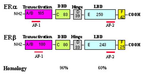
A structural comparison of human ERα and ERβ. A schematic structural comparison of human ERα and ERβ. Receptor domains are illustrated with different colored boxes, and the approximate size of each domain is indicated.
A graphic illustration of ligand-AR and ER cross-talk in the prostate cells. A graphic illustration of ligand-AR and ER cross-talk in the prostate cells. T – testosterone, E2 – estrogens, and 5αRD - 5α-reductase.
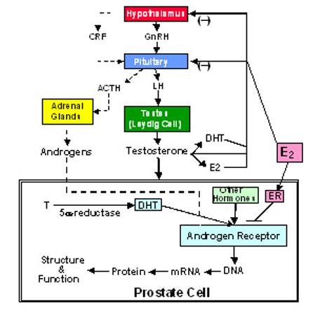
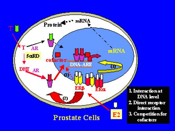
Illustration of the direct and indirect actions of estrogens in the modulation of androgen actions in the prostate cell.
Similar articles
-
The mouse as a model to investigate sex steroid metabolism in the normal and pathological prostate.J Steroid Biochem Mol Biol. 2012 Sep;131(3-5):107-21. doi: 10.1016/j.jsbmb.2011.10.009. Epub 2011 Dec 1. J Steroid Biochem Mol Biol. 2012. PMID: 22146616 Review.
-
Steroid-involved transcriptional regulation of human genes encoding prostatic acid phosphatase, prostate-specific antigen, and prostate-specific glandular kallikrein.Endocrinology. 1997 Sep;138(9):3764-70. doi: 10.1210/endo.138.9.5413. Endocrinology. 1997. PMID: 9275063
-
Steroid receptors in prostate cancer tissues and cells: pathophysiology, problems in methodology, clinical value and controversial questions.Arch Esp Urol. 1994 Mar;47(2):189-201. Arch Esp Urol. 1994. PMID: 8002681 Review.
-
Androgen receptors and the molecular basis for the action of antiadrogens in the ventral prostate.J Reprod Fertil Suppl. 1976 Sep;(24 suppl):147-62. J Reprod Fertil Suppl. 1976. PMID: 794465 Review.
-
Targeting rapid action of sex steroid receptors in breast and prostate cancers.Front Biosci (Landmark Ed). 2011 Jun 1;16(6):2224-32. doi: 10.2741/3849. Front Biosci (Landmark Ed). 2011. PMID: 21622172 Review.
Cited by
-
DHT deteriorates the progression of monocrotaline-induced pulmonary arterial hypertension: effects of endogenous and exogenous androgen.Am J Transl Res. 2019 Sep 15;11(9):5752-5763. eCollection 2019. Am J Transl Res. 2019. PMID: 31632545 Free PMC article.
-
Trans-, cis-, and dihydro-resveratrol: a comparative study.Chem Cent J. 2011 Dec 20;5:88. doi: 10.1186/1752-153X-5-88. Chem Cent J. 2011. PMID: 22185600 Free PMC article.
-
Androgen stimulates endothelial cell proliferation via an androgen receptor/VEGF/cyclin A-mediated mechanism.Am J Physiol Heart Circ Physiol. 2011 Apr;300(4):H1210-21. doi: 10.1152/ajpheart.01210.2010. Epub 2011 Jan 21. Am J Physiol Heart Circ Physiol. 2011. PMID: 21257919 Free PMC article.
-
Role of androgens in cardiovascular pathology.Vasc Health Risk Manag. 2018 Oct 15;14:283-290. doi: 10.2147/VHRM.S173259. eCollection 2018. Vasc Health Risk Manag. 2018. PMID: 30410343 Free PMC article. Review.
-
Suppression of DHT-induced paracrine stimulation of endothelial cell growth by estrogens via prostate cancer cells.Prostate. 2013 Jul;73(10):1069-81. doi: 10.1002/pros.22654. Epub 2013 Feb 19. Prostate. 2013. PMID: 23423946 Free PMC article.
References
-
- Chang C, Kokontis J, Liao S. Molecular cloning of human and rat complementary DNA encoding androgen receptors. Science. 1988;240:324–326. - PubMed
-
- Lubahn DB, Joseph DR, Sullivan PM, Willard HF, French FS, Wilson EM. Cloning of human androgen receptor complementary DNA and localization to the X chromosome. Science. 1988;240:327–330. - PubMed
-
- Quigley CA, De Bellis A, Marschke KB, el-Awady MK, Wilson EM, French FS. Androgen receptor defects: historical, clinical, and molecular perspectives. Endocr Rev. 1995;16:271–321. - PubMed
-
- Heinlein CA, Chang C. Androgen receptor (AR) co-regulators: an overview. Endocr Rev. 2002;23:175–200. - PubMed
-
- La Spada AR, Wilson EM, Lubahn DB, Harding AE, Fischbeck KH. Androgen receptor gene mutations in X-linked spinal and bulbar muscular atrophy. Nature. 1991;352:77–79. - PubMed
Grants and funding
LinkOut - more resources
Full Text Sources