Review
. 2006 Oct:213:36-47.
doi: 10.1111/j.1600-065X.2006.00436.x.

Stranger in a strange land

Affiliations
Review

Stranger in a strange land

Joan S Hunt. Immunol Rev. 2006 Oct.

Abstract

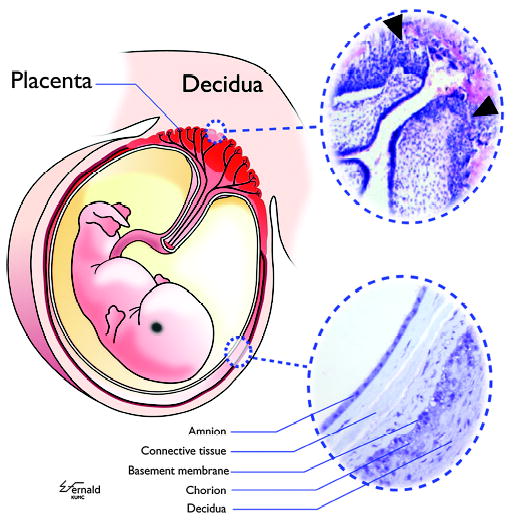
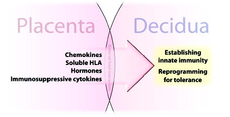
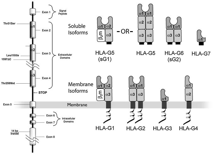
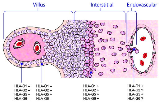
Mammalian mothers and their embryos/fetuses are almost invariably genetically different, which raises the question of how the mother's immune system is diverted so as to permit cohabitation with the 'foreign' body. Several decades of research have shown that multiple cooperative systems sanction uteroplacental immune privilege. These systems include production of several varieties of soluble immunosuppressive molecules in the uterus and the placenta and strict regulation of the molecules expressed on or by placental trophoblast cells. Trophoblast, a unique lineage without counterpart in adult tissues, is in direct contact with maternal blood and tissue. The major graft rejection-promoting molecules, human leukocyte antigens (HLAs), are tightly regulated in these cells, with none of HLA-A, HLA-B, or HLA class II antigens expressed. The HLA class Ib antigens, HLA-E, HLA-F, and HLA-G, are detectable on some subpopulations. Our studies have focused on the expression, regulation, and functions of the soluble isoforms of HLA-G, which circulate in maternal blood and are present at high levels in the pregnant uterus. These isoforms are derived from the single HLA-G gene by alternative splicing and are now known to have immunosuppressive properties. Ours and other studies indicate that soluble HLA-G proteins may comprise a unique tolerogenic system for establishing local immune privilege during pregnancy.


Figures

Fig. 1
Fig. 1. A schematic drawing of the human fetus contained within the uterus
Within the uterus, the embryo/fetus is entirely surrounded and encased by a shell of trophoblast cells present in the placenta and the chorion membrane (colored red in left figure). vCTB cells in placental villi serve as the progenitors for all differentiated trophoblast cell subpopulations, including the sTB layer, which is exposed continuously to maternal blood, and the xvCTB cells that migrate into the modified uterine endometrium, which is termed the decidua. (Upper right insert) CTB cells proliferate and migrate into the decidua (arrowheads), attaching the placental villi to the mother and facilitating certain critical physiological events required for successful pregnancy. (Lower right insert) The amnion membrane, comprising a single layer of epithelial cells, is a strong sac holding the fetus suspended in amniotic fluid. The amnion is separated from the chorion membrane CTB cells, which are derived from the migrating xvCTB cells, by connective tissue. The chorion CTB cells are directly apposed to maternal decidual cells. The inserts are photomicrographs of hematoxylin and eosin-stained placenta of 5 weeks of gestation (upper right) and an amniochorion (lower right) at termination of pregnancy. Modified from Hunt et al.(5).
Fig. 2
Fig. 2. Soluble products of the fetal placenta drive tolerance in the pregnant uterus
The fetal placenta is directly apposed and is in contact with the decidua throughout pregnancy. Placental cells, mainly vCTB and sTB within the villi, synthesize soluble substances that program maternal immune cells in the adjacent decidua. Not shown in this figure are the substances on trophoblast cell membranes that have complementary functions.
Fig. 3
Fig. 3. Multiple HLA-G proteins result from alternative splicing of the single HLA-G mRNA
(Left) The eight exons of the HLA-G gene are arranged in the same sequence as other HLA class I genes. Unlike the other HLA class I genes, the HLA-G gene is alternatively spliced to yield seven transcripts. (Right) Four messages encode membrane isoforms (HLA-G1, HLA-G2, HLA-G3, and HLA-G4), and two encode soluble isoforms (HLA-G5 and HLA-G6, also known as sG1 and sG2, respectively). A stop sequence in intron 4 results in HLA-G5 and HLA-G6, and a stop codon in intron 2 results in a third, HLA-G7. HLA-G1 and HLA-G5 produced in some but not in all types of cells associate with light chain, β2m, whereas other isoforms do not.
Fig. 4
Fig. 4. HLA-G protein expression related to column formation and invasion of the decidua
HLA-G5, a soluble glycoprotein, is the only isoform that is expressed in all subpopulations of trophoblast cells, including those in the villous (left), those in the column, and those forming the trophoblastic shell that attaches to the decidua. All tested isoforms appear to be expressed in the trophoblastic shell and to be maintained as the cells progress through the interstitum of the decidua and invade the spiral arteries.
Fig. 5
Fig. 5. An illustration of the inhibitory members of the LILR family
The inhibiting LILRs lack the arginine residue that is characteristic of the activating members of this family and have varying configurations of the extracellular portions of the molecules. LILRB1 (ILT2), LILRB2 (ILT4), and LILRB3 through B5 have long cytoplasmic tails with two to four immunoreceptor tyrosine-based inhibitory motif domains of varying sequences. Leukocytes express one or more of the LILRB receptors. Modified from Brown et al.(66).
Fig. 6
Fig. 6. Inhibitory LILR cancel activating signals
Ligation of inhibitory LILR results in phosphorylation of their ITIM domains and recruitment of p56lckand SHP-1. This causes dephosphorylation of the ITAMS that would, in the absence of the inhibitory signals, drive cell activation. HLA-G interacts with inhibitory LILR. Modified from Brown et al.(66). ITAMS, immunoreceptor tyrosine-based activating motifs; ITIM, immunoreceptor tyrosine-based inhibitory motif; SHP-1, SH2-containing protein-tyrosine-phosphatase-1.
Fig. 7
Fig. 7. Multiple conditions regulate expression of HLA-G isoforms in trophoblast cells
The vCTB cell (center) may proliferate and drive toward the maternal decidua (lower portion of figure). Acquisition of higher levels of isoform-specific HLA-G mRNAs occurs as a consequence of low oxygen levels in the decidua. Forces driving protein expression in these cells are not known. Alternatively, vCTB cells may differentiate into sTB (upper portion of figure). Epidermal growth factor appears to increase HLA-G5 and reduce HLA-G6 mRNA in these cells and to promote exclusive expression of HLA-G5 in the differentiated syncytium.
Fig. 8
Fig. 8. Substrate influences expression of HLA-G5 but not HLA-G6 in vCTB cells
vCTB cells grown on uncoated plates for 48 h show granular HLA-G5 in cell cytoplasm (arrowheads), whereas cells grown on plates coated with type IV collagen have large intracellular inclusions of HLA-G5 (arrowheads). HLA-G2/G6 appeared to be unchanged. FcR binding of the isotype-specific control monoclonal antibody was more frequent in vCTB cells grown on uncoated plates (arrow) than on coated plates. Original magnifications ×200.
Fig. 9
Fig. 9. Immunofluorescent staining for syntaxin-4, an intracellular transport protein (SNARE), in mononuclear phagocyte-like THP-1 cells
This SNARE is present in the monocyte cell line THP-1 following differentiation with phorbol myristate acetate for 24 h and activation with IFNγ for an additional 24 h. Preliminary experiments suggest that proteins such as syntaxin-4 are altered in these cells following treatment with rHLA-G5, which could affect secretion of cytokines. Blue signal, DAPI nuclear stain; green signal, anti-syntaxin-4. The insert shows negative results with the isotype control for anti-syntaxin-4. Original magnifications ×250.

References

    1. Heinlein RA. New York: GP Putnam’s Sons; 1961. Stranger in a Strange Land.
    1. Burton GJ, Hempstock J, Jauniaux E. Oxygen, early embryonic metabolism and free radical-mediated embryopathies. Reprod Biomed Online. 2002;6:84–96. - PubMed
    1. Bulmer JN, Pace D, Ritson A. Immunoregulatory cells in human decidua: morphology, immunohistochemistry and function. Reprod Nutr Dev. 1988;28:1599–1613. - PubMed
    1. Rossant J, Cross JC. Placental development: lessons from mouse mutants. Nat Rev Genet. 2001;2:538–548. - PubMed
    1. Hunt JS, Petroff MG, McIntire RH, Ober C. HLA-G and immune tolerance in pregnancy. FASEB J. 2005;19:681–693. - PubMed

Publication types