Review
. 2006 Oct-Dec;27(5-6):483-94.
doi: 10.1016/j.mam.2006.08.004. Epub 2006 Sep 14.

Autophagy in organelle homeostasis: peroxisome turnover

Affiliations
Review

Autophagy in organelle homeostasis: peroxisome turnover

Iryna Monastyrska et al. Mol Aspects Med. 2006 Oct-Dec.

Abstract

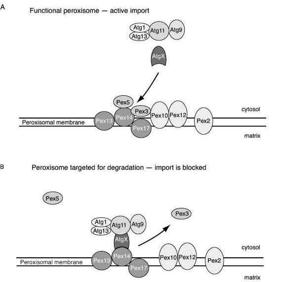
When cells are confronted with an insufficient supply of nutrients in their extracellular fluid, they may begin to cannibalize some of their internal proteins as well as whole organelles for reuse in the synthesis of new components. This process is termed autophagy and it involves the formation of a double-membrane structure within the cell, which encloses the material to be degraded into a vesicle called an autophagosome. The autophagosome subsequently fuses with a lysosome/vacuole whose hydrolytic enzymes degrade the sequestered organelle. Degradation of peroxisomes is a specific type of autophagy, which occurs in a selective manner and has been mostly studied in yeast. Recently, it was reported that a similar selective process of autophagy occurs in mammalian cells with proliferated peroxisomes. Here we discuss characteristics of the autophagy of peroxisomes in mammalian cells and present a comprehensive model of their likely mechanism of degradation on the basis of known and common elements from other systems.


Figures

Figure 1
Figure 1
Selective degradation of peroxisomes via macropexophagy and micropexophagy. Morphological events. Macropexophagy starts at a single peroxisome. This process involves targeting of peroxisome for initial membrane nucleation, sequestration–formation of autophagosomes–and hydrolytic degradation inside the vacuole/lysosome. In contrast, during micropexophagy a cluster of peroxisomes without prior sequestration is directly engulfed by vacuolar protrusions. These peroxisomes are released and degraded in the vacuolar lumen similar to the last stages of macropexophagy. P, peroxisome.
Figure 2
Figure 2
Targeting of peroxisome for degradation. Model of molecular mechanism underlying initiation of pexophagy. A. By docking to the peroxisomal membrane and subsequent translocation, Pex5 shuttles between the cytosol and matrix, taking newly synthesized peroxisomal proteins into the lumen. The peroxisomal membrane protein Pex3 bridges the docking peroxisomal complex (Pex13, Pex14 and Pex17) and the putative translocation site (Pex12, Pex10 and Pex2). In this way peroxisome matrix proteins are imported and the peroxisome is functional. At this stage Pex3 presumably shields Pex14. B. When peroxisomes are no longer functional, the docking and translocation complexes are apart. Pex3 is removed, thus exposing Pex14 for targeting by a hypothetical receptor (AtgX). Via AtgX and Atg11 the peroxisome is connected to the other Atg components, allowing autophagic sequestration to occur.
Figure 3
Figure 3
Molecular mechanism of autophagosome formation. A single import-incompetent peroxisome is targeted for degradation. Peroxisomal membrane complex modifications from import–competent to incompetent allow docking of sequestering membranes to the peroxisome. This process occurs through multiple sequential steps carried out by Atg proteins. The Atg1-Atg13 and PtdIns 3-kinase complexes are involved in forming a scaffold that allows for the initiation of autophagosome formation. Atg8 and Atg9 are involved in the building of membranes by bringing lipids by recruitment and subsequent release. Atg9 cycles to the mitochondria, which possibly is the donor of lipids. Atg8 is involved in the recruitment of the Atg12–Atg5-Atg16 complex, which forms a coat that might be necessary for membrane curvature. Prior to completion of the autophagosome most of the Atg proteins dissociate from the membrane.

References

    1. Agne B, Meindl NM, Niederhoff K, Einwachter H, Rehling P, Sickmann A, Meyer HE, Girzalsky W, Kunau WH. Pex8p: An intraperioxisomal organizer of the peroxisomal import machinery. Mol. Cell. 2003;11:635–646. - PubMed
    1. Aplin A, Jasionowski T, Tuttle DL, Lenk SE, Dunn WA. Cytoskeletal elements are required for the formation and maturation of autophagic vacuoles. J. Cell. Physiol. 1992;152:458–466. - PubMed
    1. Baerends RJS, Faber KN, Kram AM, Kiel JAKW, van der Klei I, Veenhuis M. A stretch of positively charged amino acids at the N terminus of Hansenula polymorpha Pex3p is involved in incorporation of the protein into the peroxisomal membrane. J Biol Chem. 2000;275:9986–9995. - PubMed
    1. Bellu AR, Komori M, van der Klei I, Kiel JAKW, Veenhuis M. Peroxisome biogenesis and selective degradation converge at Pex14p. J Biol Chem. 2001;276:44570–44574. - PubMed
    1. Bellu AR, Salomons FA, Kiel JAKW, Veenhuis M, van der Klei I. Removal of Pex3p is an important initial stage in selective peroxisome degradation in Hansenula polymorpha. J Biol Chem. 2002;277:42875–42880. - PubMed

Publication types

LinkOut - more resources