Review
. 2006 Dec;18(6):375-86.
doi: 10.1016/j.smim.2006.07.009. Epub 2006 Sep 14.

Mendelian resistance to human norovirus infections

Affiliations
Review

Mendelian resistance to human norovirus infections

Jacques Le Pendu et al. Semin Immunol. 2006 Dec.

Abstract

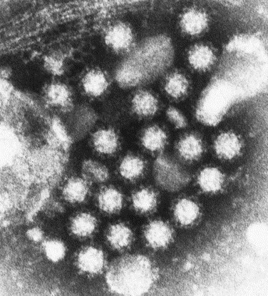
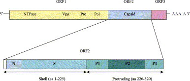
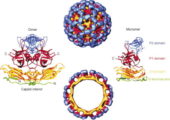
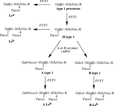
Noroviruses have emerged as a major cause of acute gastroenteritis in humans of all ages. Despite high infectivity of the virus and lack of long-term immunity, volunteer and authentic studies has suggested the existence of inherited protective factors. Recent studies have shown that histo-blood group antigens (HBGAs) and in particular secretor status controlled by the alpha1,2fucosyltransferase FUT2 gene determine susceptibility to norovirus infections, with nonsecretors (FUT2-/-), representing 20% of Europeans, being highly resistant to symptomatic infections with major strains of norovirus. Moreover, the capsid protein from distinct strains shows different HBGA specificities, suggesting a host-pathogen co-evolution driven by carbohydrate-protein interactions.


Figures

Fig. 1
Fig. 1
Electron microscopy of noroviruses (29 nm) from clinical sample. Virus particles visualized by negative staining (phosphotungstic acid).
Fig. 2
Fig. 2
Schematic representation of the noroviral genome, which is divided into three open reading frames: ORF1–3. ORF1 encodes four nonstructural proteins, namely NTPase (nucleoside triphosphate) VPg, proteinase and polymerase. ORF2 encodes the capsid of the virus and ORF3 encodes a minor structural protein. Bottom part: Schematic illustration of ORF2, encoding the capsid of the norovirus. N is the NH2-terminal arm of ORF2 and S refers to the inner shell; the most conserved part of the capsid. P1 and P2 are the protruding parts, of which P2 is the most variable and exposed.
Fig. 3
Fig. 3
The caliciviruses are divided into four different genera: Lagovirus, Vesivirus, Sapovirus and Norovirus of which the last two can infect humans. Noroviruses is further subdivided into five distinct genogroups, GGI–GGV.
Fig. 4
Fig. 4
Neighbor-joining phylogenetic tree of norovirus genogroups based on the complete capsid region. Genogroups (GG) and genotypes (numbers after GG) are indicated. Percentage bootstrap values are given at branch nodes, and the number of substitutions per site is indicated by the scale bar.
Fig. 5
Fig. 5
Cryo-electron microscopy (22 Å) and X-ray crystallography (3.4 Å) of Norwalk virus-like particles. Surface representation (top) and cross-section. The virus-like particles have 90 dimers of the capsid (left ribbon diagram) in a T = 3 icosahedral symmetry. The capsid protein is divided into an N-terminal region (green) facing the interior of the VLP, a shell domain (S-domain, yellow) that constitutes the surface of the virus, and a protruding domain (P-domain) that emanates from the S-domain surface. The P-domain is divided into P1 and P2 (red and blue, respectively) with the P2-domain at the most distal surface of the particle. With permission from Refs. , , .
Fig. 6
Fig. 6
Biosynthesis of ABH and Lewis histo-blood group antigens (HBGAs) proceeds by stepwise addition of monosaccharides to precursor structures. The figure shows the example of the so-called type 1 precursor Galβ1-3GlcNAc. H antigen synthesis is under dependence of the FUT2 gene encoding an α1,2fucosyltransferase which attaches a fucose residue in α1,2 linkage to the terminal galactose of the precursor. Inactivating mutations of FUT2 are responsible for a phenotype called “nonsecretor”, characterized by the lack of α1,2-linked fucose-containing HBGAs on many epithelial cells and in secretions such as saliva. Synthesis of the A and B antigens requires the previous synthesis of the H antigen which will be used as an acceptor substrate by the A and B enzymes encoded by the A and B alleles at the ABO locus. These enzymes will add an N-acetylgalactosamine or a galactose in α1,3 linkage on the galactose residue of the H antigen to give the A and B antigens, respectively. O alleles at the ABO locus correspond to inactivated alleles that cannot encode for a functional glycosyltransferase. The Lewis antigens are synthesized thanks to α3/4fucosyltransferases that attach a fucose residue on the N-acetylglucosamine of the precursor. In the case of type 1 precursor, this fucose residue is in α1,4 linkage and its addition gives the Lewis a (Lea) and Lewis b (Leb) antigens. The gene mainly responsible for their synthesis in epithelia is FUT3 for which, similar to FUT2 and ABO, inactivated alleles are known. Individual homozygous for inactivated FUT3 alleles (FUT3−/−) are essentially devoid of α4-linked fucose on their epithelial cells and in saliva and their phenotype is usually referred to as “Lewis negative”. Enzymes are in italics and the antigens are named in bold. Gal: galactose; GlcNAc: N-acetylglucosamine; Fuc: fucose; GalNAc: N-acetylgalactosamine.
Fig. 7
Fig. 7
Human infectivity spectrum deduced from the carbohydrate-binding specificities of noroviruses strains. (A) The human population is divided into subgroups depending upon the combined polymorphisms at the FUT2, ABO and FUT3 loci. For simplicity, the Lewis negative phenotype (FUT3−/−) is not shown within the Secretor (FUT2+) subgroup. Phenotypic subdivisions are shown in the figure according to approximate frequencies observed in Western Europe. Significant differences in allelic frequencies at each locus can be observed in different parts of the world. Eight types of norovirus strains are depicted according to their ability to bind distinct HBGAs structures. Type 1 corresponds to the VA387 and Grimsby strains, type 2 to NV, type 3 to Mexico, type 4 to MOH, type 5 to SMV, type 6 to BUDS, type 7 to VA207 and Boxer, type 8 to OIF. Since the presence of a given carbohydrate depends upon the genetic polymorphism, the spectrum of individuals that one type of strain can recognize is limited to a fraction of the population. Cross-reactivities may occur as exemplified by strains of the types 2, 3 and 7 that can extend their host recognition beyond their main target phenotype. In the case of type 2 strains exemplified by the Norwalk strain, blood group B individuals are less well recognized. They are less likely infected and if so, they can remain asymptomatic. In the case of type 7 strains, although the main targets should be nonsecretors/Lewis positive individuals (FUT2−/FUT3+), some blood group O Secretors (FUT2+) should be infected providing they are Lewis positive (FUT3+). (B) Monosaccharides units at three positions can be subject to a polymorphism and the eight types of strains depicted in A can be explained by the preferential targeting of the monosaccharide residue at one of these three variable positions. The fucose residues (circles) are present or absent depending upon the FUT2 and FUT3 genes polymorphisms (blue and green respectively). The N-acetylgalactosamine or galactose (brown triangle) can be either present or absent depending upon the ABO gene polymorphism. Some strains will target mainly the α1,2-linked fucose (types 1 and 2), others will target mainly the N-acetylgalactosamine or the galactose residue (types 3–6), while others yet will target mainly the α1,4-linked fucose (types 7 and 8). These major specificities are figured by dark colors of arc of circles. Strain binding specificity can extend more or less beyond the main target epitope (arc of circles lighter colors). The figures correspond to interpretation of data obtained by different methods and reported in Refs. , , , , .

Similar articles

Cited by

References

    1. Casanova J.-L., Abel L. Inborn errors of immunity to infection: the rule rather than the exception. J Exp Med. 2005;202:197–201. - PMC - PubMed
    1. Ohtsuka N., Taguchi F. Mouse susceptibility to mouse hepatitis virus infection is linked to viral receptor genotype. J Virol. 1997;71:8860–8863. - PMC - PubMed
    1. Brown K.E., Hibbs J.R., Gallinella G. Resistance to parvovirus B19 infection due to lack of virus receptor (erythrocyte P antigen) N Engl J Med. 1994;330:1192–1196. - PubMed
    1. Kindberg E., Hejdeman B., Bratt G. A nonsense mutation (428G → A) in the fucosyltransferase FUT2 gene affects the progression of HIV-1 infection. AIDS. 2006;20:685–689. - PubMed
    1. Glass W.G., Lim J.K., Cholera R., Pletnev A.G., Gao J.L., Murphy P.M. Chemokine receptor CCR5 promotes leukocyte trafficking to the brain and survival in West Nile virus infection. J Exp Med. 2005;202:1087–1098. - PMC - PubMed

Publication types