. 2006 Oct;80(19):9455-64.
doi: 10.1128/JVI.01149-06.

Vaccinia virus F9 virion membrane protein is required for entry but not virus assembly, in contrast to the related L1 protein

Affiliations

Vaccinia virus F9 virion membrane protein is required for entry but not virus assembly, in contrast to the related L1 protein

Erica Brown et al. J Virol. 2006 Oct.

Abstract

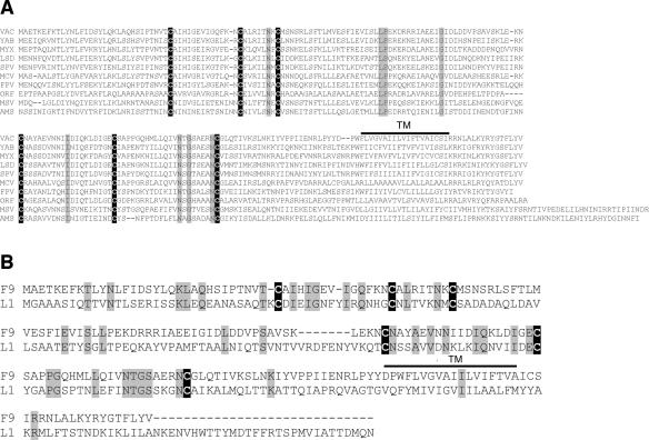
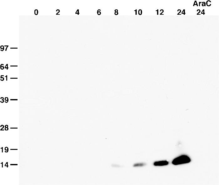
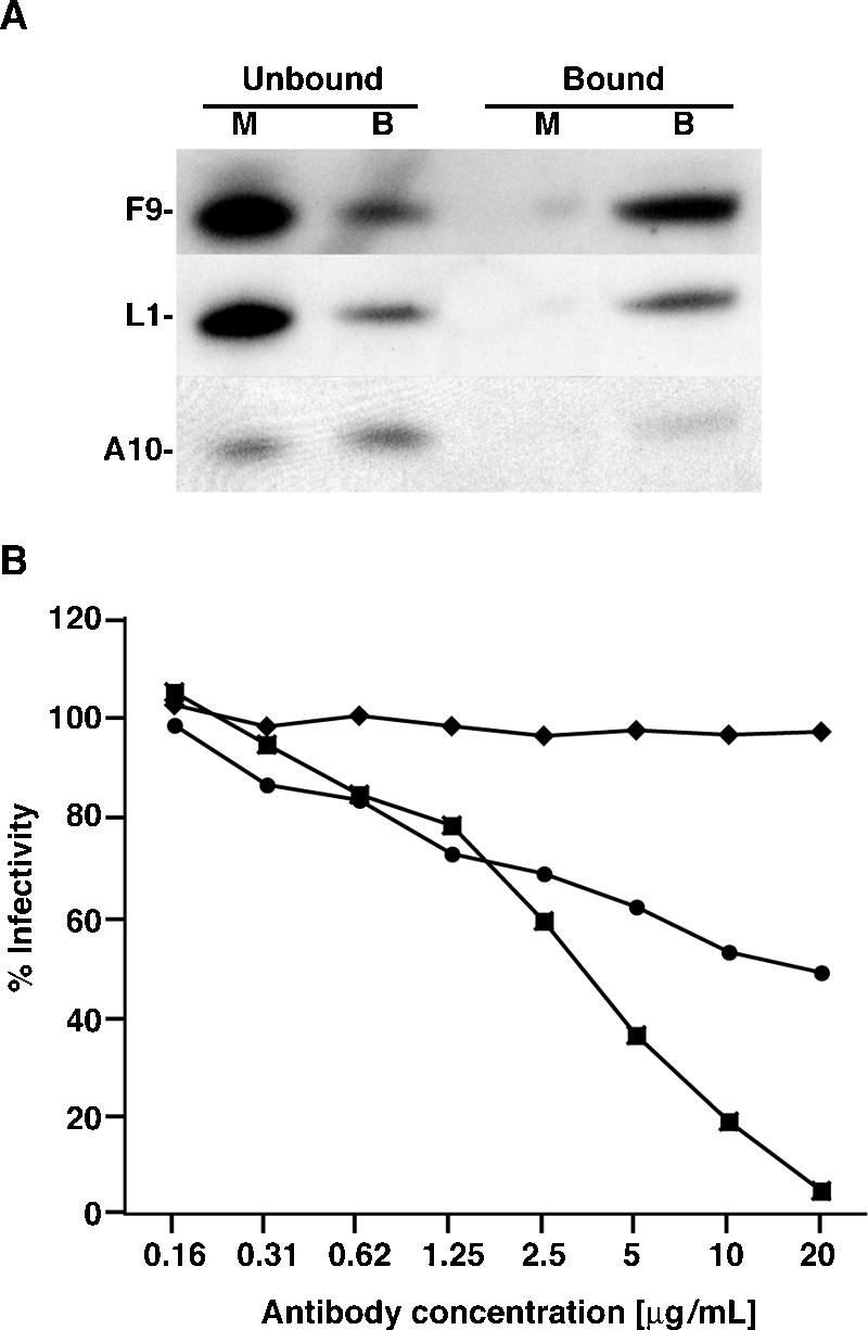
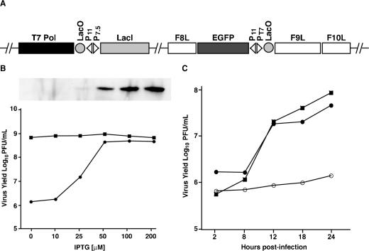
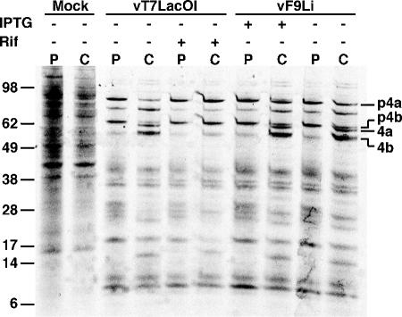
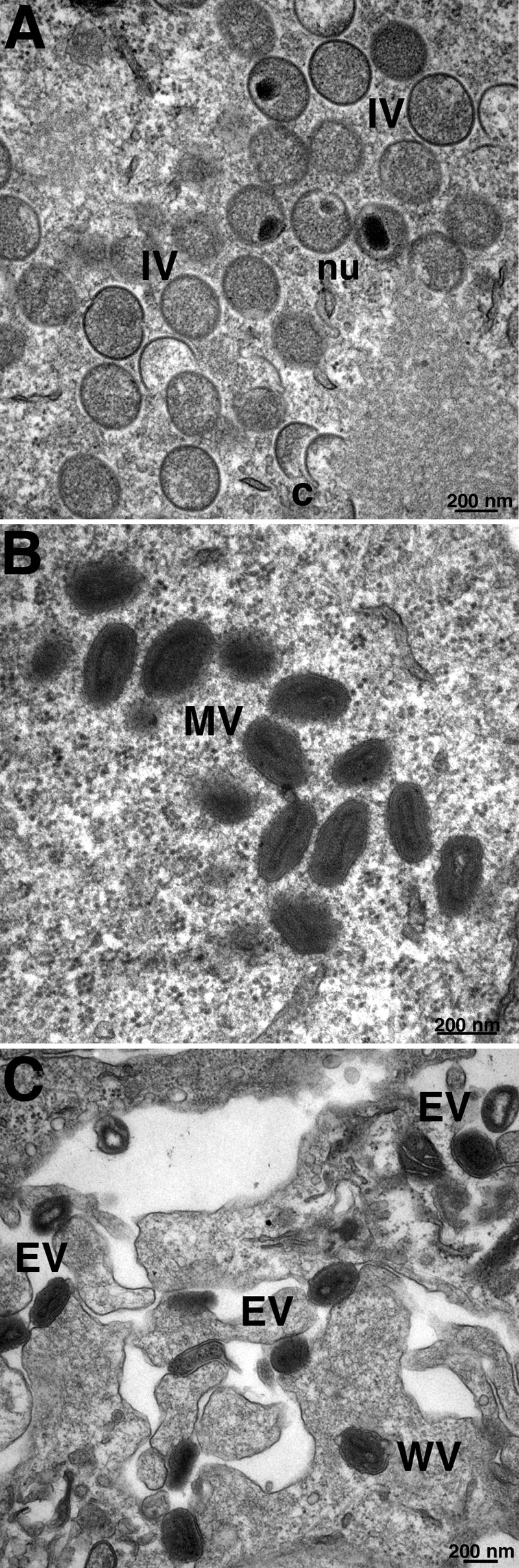
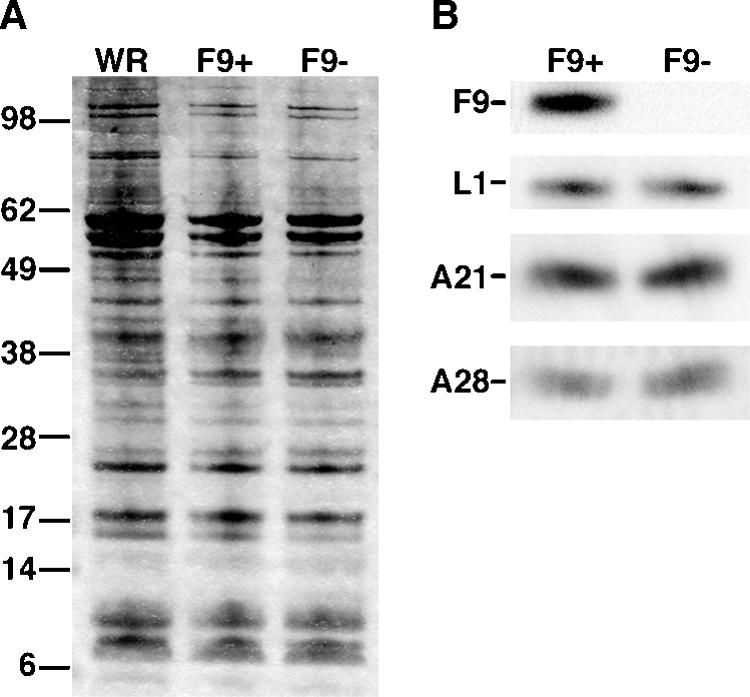
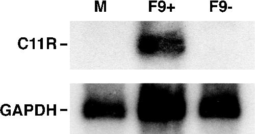
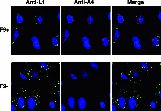
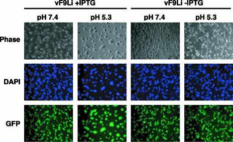
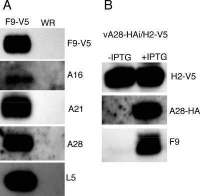
All sequenced poxviruses encode orthologs of the vaccinia virus L1 and F9 proteins, which are structurally similar and share about 20% amino acid identity. We found that F9 further resembles L1 as both proteins are membrane components of the mature virion with similar topologies and induce neutralizing antibodies. In addition, a recombinant vaccinia virus that inducibly expresses F9, like a previously described L1 mutant, had a conditional-lethal phenotype: plaque formation and replication of infectious virus were dependent on added inducer. However, only immature virus particles are made when L1 is repressed, whereas normal-looking intracellular and extracellular virions formed in the absence of F9. Except for the lack of F9, the polypeptide components of such virions were indistinguishable from those of wild-type virus. These F9-deficient virions bound to cells, but their cores did not penetrate into the cytoplasm. Furthermore, cells infected with F9-negative virions did not fuse after a brief low-pH treatment, as did cells infected with virus made in the presence of inducer. In these respects, the phenotype associated with F9 deficiency was identical to that produced by the lack of individual components of a previously described poxvirus entry/fusion complex. Moreover, F9 interacted with proteins of that complex, supporting a related role. Thus, despite the structural relationships of L1 and F9, the two proteins have distinct functions in assembly and entry, respectively.


Figures

FIG. 1.
FIG. 1.
F9 is conserved throughout the Poxviridae. (A) Multiple amino acid sequence alignment of VACV F9 and orthologs from representatives of each poxvirus genus. The bar denotes the predicted transmembrane (TM) domain. Identical amino acids are shaded gray; invariant cysteines are white on a black background. Abbreviations: VAC, vaccinia virus (Orthopoxvirus); YAB, Yaba monkey tumor virus (Yatapoxvirus); MYX, myxoma virus (Leporipoxvirus); LSD, lumpy skin disease virus (Capripoxvirus); SPV, swinepox virus (Suipoxvirus); MCV, molluscum contagiosum virus (Molluscipoxvirus); FPV, fowlpox virus (Avipoxvirus); ORF, ORF virus (Parapoxvirus); MSV, Melanoplus sanguinipes entomopoxvirus (Entomopoxvirus B); AMS, Amsacta moorei entomopoxvirus (Entomopoxvirus B). (B) Sequence alignment of VACV WR F9 and L1 proteins. Identical amino acids are denoted as in panel A.
FIG. 2.
FIG. 2.
Kinetics of F9 expression. BS-C-1 cells were infected with VACV for the hours indicated at the top of the figure, harvested in the presence of N-ethylmaleimide, and analyzed by nonreducing SDS-PAGE and Western blotting with a rabbit polyclonal antibody specific for F9 followed by horseradish peroxidase-conjugated anti-rabbit antibody. One infection was performed in the presence of AraC, and cells were harvested at 24 h. Bands were detected by chemiluminescence. Numbers at left show molecular masses in kilodaltons.
FIG. 3.
FIG. 3.
F9 is an MV membrane protein. (A) Biotinylation of MV membrane proteins. Purified VACV virions were mock treated (lanes M) or treated with 1 mg/ml of sulfo-NHS-SS-biotin (lanes B) for 30 min at 4°C. Virions were lysed with 0.5% SDS, and biotinylated proteins were isolated by incubation with NutrAvidin resin. Bound proteins were eluted with 0.5% SDS containing 50 mM DTT. Western blot analysis was performed using antibodies specific for F9, L1, and A10. (B) MV neutralization (11). Purified WR-NPsiinfeklEGFP MVs were mixed with various concentrations of polyclonal antibody to F9 (circles), L1 (squares), or A33 (diamonds) for 1 h at 37°C. The mixture was then added to HeLa cells in the presence of AraC and incubated for 16 h. EGFP-expressing cells were quantified by flow cytometry. Data are presented as the percentage of EGFP-expressing cells normalized to cells incubated with virus that was mock treated with antibody.
FIG. 4.
FIG. 4.
F9L is essential for replication of infectious virus. (A) Diagram of vF9Li. A segment of the viral genome shows the open reading frames for the bacteriophage T7 RNA polymerase (T7 Pol), E. coli lac repressor (LacI), EGFP, VACV F9L, F8L, and F10L. Also indicated are the E. coli Lac operator (LacO) and the VACV promoters P11 and P7.5 and T7 promoter PT7. (B) Effect of IPTG concentration on virus yield and F9 expression. BS-C-1 cells were infected with 10 PFU per cell of vT7LacOI (squares) or vF9Li (circles) in the presence of indicated concentrations of IPTG. At 24 h postinfection, cells were harvested, subjected to three cycles of freezing-thawing, and sonicated. Virus yields were determined by plaque assay on BS-C-1 cells in the presence of 100 μM IPTG. The inset shows F9 protein at 24 h as a function of IPTG concentration assayed by Western blotting with anti-F9 antibody. (C) One-step virus growth. BS-C-1 cells were infected with 10 PFU per cell of vT7LacOI (filled squares) or vF9Li in the presence (filled circles) or absence (open circles) of 100 μM IPTG. At the indicated times, virus was harvested from infected cells and plaque assayed as described above.
FIG. 5.
FIG. 5.
Processing of viral core proteins. BS-C-1 cells were infected with 5 PFU per cell of vF9Li in the presence or absence of IPTG or vT7LacOI in the presence or absence of 100 μg/ml of rifampin. After 11.5 h, cells were incubated in methionine- and cysteine-deficient medium for 30 min and then with 100 μCi of a [35S]methionine and [35S]cysteine mixture for 30 min. The cells were washed and harvested either immediately after the pulse (P) or after incubation in unlabeled medium for an additional 12-h chase (C). Cell lysates were analyzed by SDS-PAGE and autoradiography. Core precursor proteins p4a and p4b as well as processed proteins 4a and 4b are indicated. Positions of marker proteins are shown at the left with masses in kilodaltons.
FIG. 6.
FIG. 6.
Electron microscopy of cells infected with vF9Li in the absence of IPTG. BS-C-1 cells were infected with vF9Li at a multiplicity of 4 PFU per cell in the absence of IPTG. After 20 h, the cells were processed for transmission electron microscopy as previously described (41). (A) Crescents (c) and IVs with visible nucleoids (nu). (B) MVs. (C) Wrapped virions (WV) and EVs. Bars, 200 nm.
FIG. 7.
FIG. 7.
Polypeptide composition of purified F9+ and F9 virions. Purified MVs were obtained by sedimentation through two successive 36% sucrose cushions and a 25 to 40% sucrose gradient. Equal numbers of VACV WR, F9+, and F9 virions were solubilized with SDS and separated by SDS-PAGE in 4 to 12% polyacrylamide Bis-Tris gels. (A) Silver stain. The masses in kilodaltons are indicated on the left. (B) Western blot probed with anti-F9, anti-L1, anti-A21, and anti-A28 antibodies as indicated.
FIG. 8.
FIG. 8.
Early transcription in vF9Li-infected cells. BS-C-1 cells were either mock infected (M) or infected with an equal number of purified F9+ or F9 MV particles in the presence of AraC. At 3 h after infection, total RNA was harvested and a Northern blot assay was performed using a [32P]dCMP-labeled DNA probe for VACV early C11R and cellular glyceraldehyde-3-phosphate dehydrogenase (GAPDH) mRNA. An autoradiogram is shown.
FIG. 9.
FIG. 9.
Cell binding and penetration assay. HeLa cells were incubated with 10 PFU of purified F9+ virions or the same number of F9 virions at 4°C for 1 h and then shifted to 37°C for 2 h. Subsequently, cells were fixed, permeabilized, and stained for indirect immunofluorescence microscopy. The L1 MV membrane protein was stained with a mouse monoclonal anti-L1 antibody and a goat anti-mouse secondary antibody conjugated to Alexa Fluor 488 (green). The A4 core protein was stained with a rabbit primary antibody and a goat anti-rabbit IgG secondary antibody conjugated to Alexa Fluor 568 (red). Cellular DNA was stained with DAPI (blue).
FIG. 10.
FIG. 10.
Low-pH-triggered cell-cell fusion assays. BS-C-1 cells were infected with vF9Li in the presence or absence of IPTG. At 18 h after infection, the cells were treated for 5 min with pH 7.4 or pH 5.3 buffer at 37°C and then returned to normal medium. After 18 h, the cells were fixed, permeabilized, and stained with DAPI. Cells were visualized by phase-contrast microscopy, while DNA (blue) and GFP (green) staining were visualized by fluorescence microscopy.
FIG. 11.
FIG. 11.
Interaction of F9 with the entry/fusion complex. (A) BS-C-1 cells were infected with vF9-V5 or VACV WR. After 24 h, the cells were disrupted with a Dounce homogenizer and postnuclear supernatants were centrifuged at 100,000 × g for 1 h. The pellet was extracted with PBS-NP-40, and the soluble fraction was immunopurified on agarose beads conjugated to V5 antibody. Proteins were resolved by SDS-PAGE, transferred to a nitrocellulose membrane, and incubated with anti-V5 antibody conjugated to peroxidase and primary rabbit antibodies to the A16, A21, A28, and L5 proteins followed by peroxidase-conjugated secondary antibody to rabbit IgG. (B) BS-C-1 cells were infected with vA28-HAi/H2-V5 in the presence or absence of IPTG. The NP-40-PBS-soluble extract of the membrane-enriched fraction was immunopurified on agarose beads conjugated to V5 antibody and resolved by SDS-PAGE. Proteins were transferred to a nitrocellulose membrane and detected by Western blotting with peroxidase-conjugated anti-V5 or anti-HA antibody or with anti-F9 rabbit antibody followed by peroxidase-conjugated anti-rabbit IgG.

References

    1. Alcami, A. 2003. Viral mimicry of cytokines, chemokines and their receptors. Nat. Rev. Immunol. 3:36-50. - PubMed
    1. Aldaz-Carroll, L., J. C. Whitbeck, M. Ponce de Leon, H. Lou, L. K. Pannell, J. Lebowitz, C. Fogg, C. White, B. Moss, G. H. Cohen, and R. J. Eisenberg. 2005. Physical and immunological characterization of a recombinant secreted form of the membrane protein encoded by the vaccinia virus L1R gene. Virology 341:59-71. - PubMed
    1. Alexander, W. A., B. Moss, and T. R. Fuerst. 1992. Regulated expression of foreign genes in vaccinia virus under the control of bacteriophage T7 RNA polymerase and the Escherichia coli lac repressor. J. Virol. 66:2934-2942. - PMC - PubMed
    1. Armstrong, J. A., D. H. Metz, and M. R. Young. 1973. The mode of entry of vaccinia virus into L cells. J. Gen. Virol. 21:533-537. - PubMed
    1. Broyles, S. S. 2003. Vaccinia virus transcription. J. Gen. Virol. 84:2293-2303. - PubMed

Publication types

LinkOut - more resources