. 2006 Oct;80(19):9481-96.
doi: 10.1128/JVI.00533-06.

Essential functions of the unique N-terminal region of the varicella-zoster virus glycoprotein E ectodomain in viral replication and in the pathogenesis of skin infection

Affiliations

Essential functions of the unique N-terminal region of the varicella-zoster virus glycoprotein E ectodomain in viral replication and in the pathogenesis of skin infection

Barbara Berarducci et al. J Virol. 2006 Oct.

Abstract

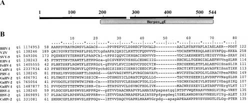
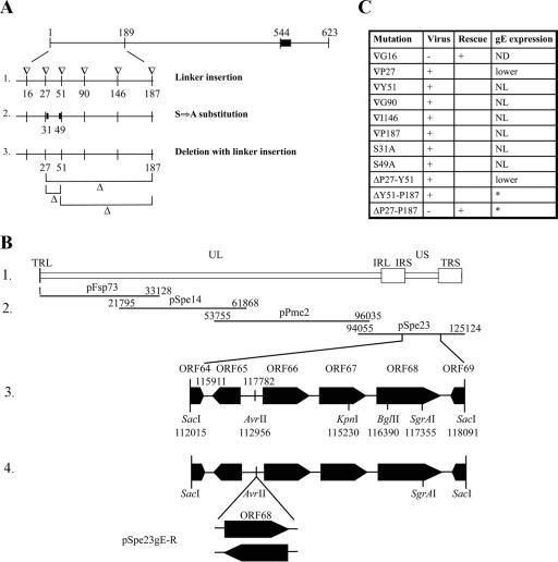
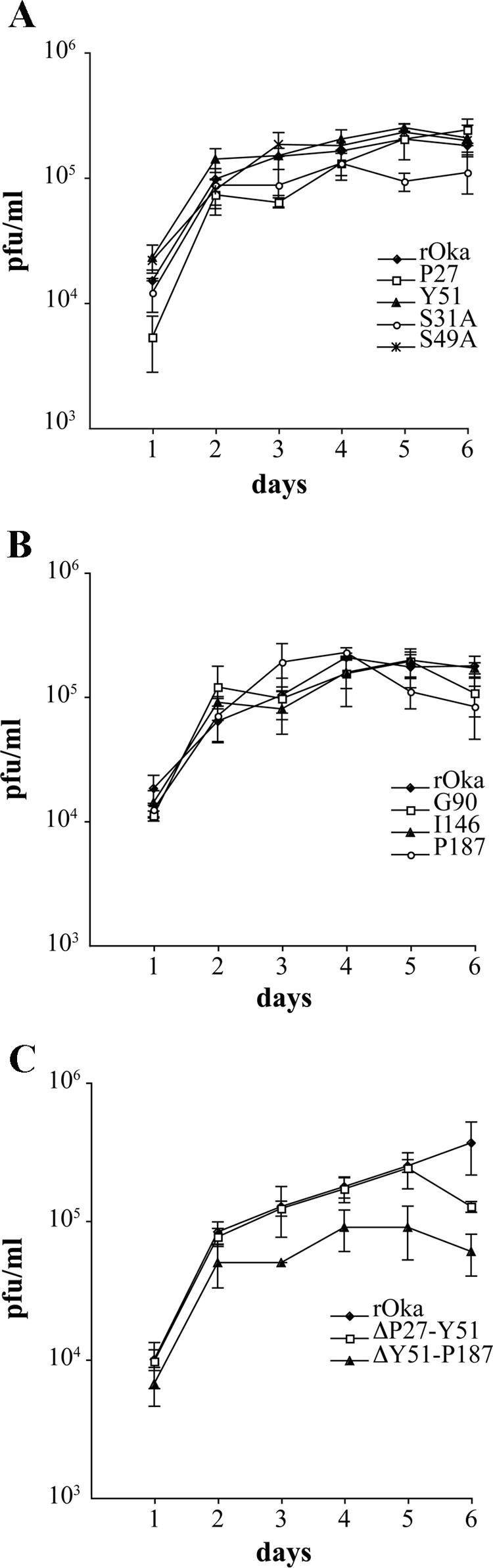
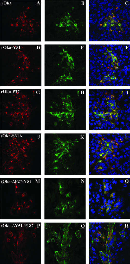
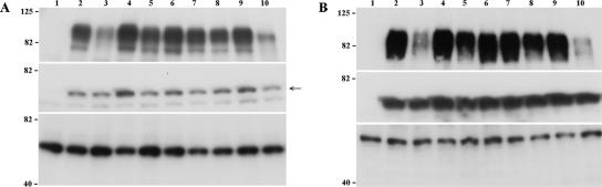
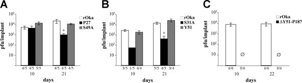
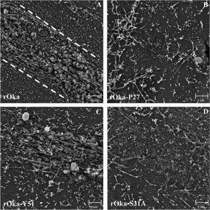
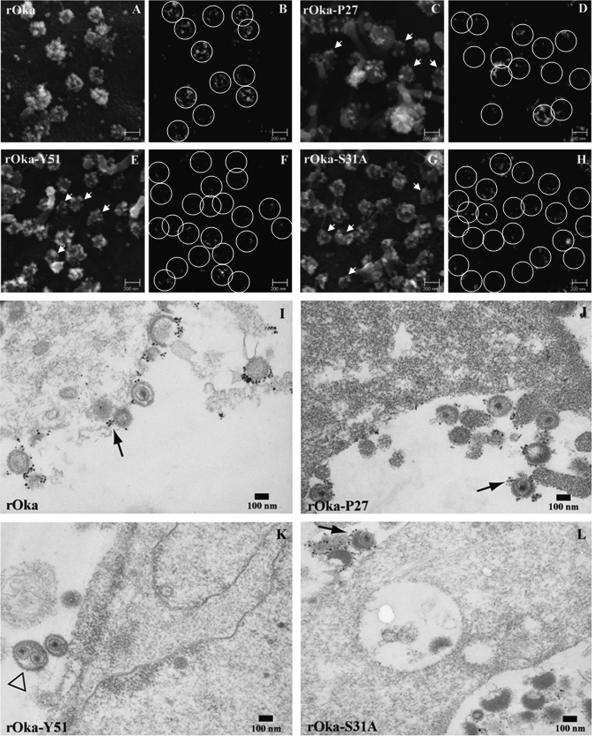
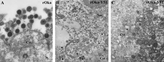
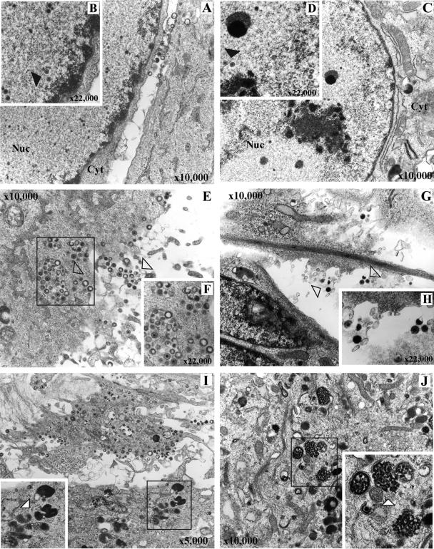
Varicella-zoster virus (VZV) glycoprotein E (gE) is a multifunctional protein important for cell-cell spread, envelopment, and possibly entry. In contrast to other alphaherpesviruses, gE is essential for VZV replication. Interestingly, the N-terminal region of gE, comprised of amino acids 1 to 188, was shown not to be conserved in the other alphaherpesviruses by bioinformatics analysis. Mutational analysis was performed to investigate the functions associated with this unique gE N-terminal region. Linker insertions, serine-to-alanine mutations, and deletions were introduced in the gE N-terminal region in the VZV genome, and the effects of these mutations on virus replication and cell-cell spread, gE trafficking and localization, virion formation, and replication in vivo in the skin were analyzed. In summary, mutagenesis of the gE N-terminal region identified a new functional region in the VZV gE ectodomain essential for cell-cell spread and the pathogenesis of VZV skin tropism and demonstrated that different subdomains of the unique N-terminal region had specific roles in viral replication, cell-cell spread, and secondary envelopment.


Figures

FIG. 1.
FIG. 1.
Identification of the conserved domain in VZV gE. The 544-amino-acid ectodomain of VZV gE was analyzed for conserved domains with the “Conserved Domains Databases” using the Pfam database (32). (A) VZV gE ectodomain of amino acids 1 to 544 and the position of the conserved domain. (B) Alignment of VZV gE and gE in the other alphaherpesviruses. The first 80 amino acids of the alignment are shown. Numbers on the left indicate the amino acid from which the conserved domain starts; numbers at the far left are the accession numbers. BHV, bovine herpesvirus; VZV, varicella-zoster virus; SVV, simian varicella virus; EHV, equine herpesvirus; FeHV, feline herpesvirus; CaHV, canine herpesvirus; PRV, pseudorabiesvirus; GaHV, gallid herpesvirus; HSV, herpes simplex virus; CeHV, SA8.
FIG. 2.
FIG. 2.
gE mutagenesis. (A) Schematic representation of the gE mutations. The gE glycoprotein and the positions of the mutations are represented. The position of the linker insertions (▿, line 1), serine-to-alanine alterations (thick bar, line 2), and deletions (Δ, line 3) are indicated. Amino acids are numbered from the N terminus to the C terminus of the gE protein. The black box represents the transmembrane domain. (B) Schema of the cosmid system. Line 1, schematic representation of the VZV genome; line 2, diagram of the overlapping cosmids containing the VZV genome (parental Oka); line 3, the SacI fragment from the pSpe23 cosmid containing the ORF68 gene; line 4, insertion of the gE rescue cassette in the AvrII site. pSpe23gE-R indicates the rescued gE mutants. (C) Summary of the results from gE mutagenesis. Recovery of recombinant virus, rescue of the mutation with lethal phenotype, and level of gE expression are indicated for each mutation. ND, not done; NL, normal. An asterisk indicates that the expression of mutant ΔY51-P187 and ΔP27-P187 gE was analyzed by immunofluorescence only (see Fig. 3, 4, and 6P to R). TRL, terminal repeat long; UL, unique long region; IRL, internal repeat long; IRS, inverted repeat short; US, unique short region; TRS, terminal repeat short.
FIG. 3.
FIG. 3.
Localization of mutant gE ΔP27-P187 in melanoma cells. Melanoma cells were cotransfected with wild-type gE (A to C and G to I) or mutant ΔP27-P187 gE (D to F and J to L) and gI, and they were analyzed 48 h posttransfection. Transfected cells were fixed and permeabilized (A to F) or fixed without permeabilization to detect the staining at the plasma membrane (G to L). gE (red) was detected with MAb 7G8, which recognizes the C terminus of the protein; gI (green) was detected with an anti-rabbit antibody against gI; nuclei were stained with 4′,6′-diamidino-2-phenylindole (blue). Magnification, ×63. wt, wild type.
FIG. 4.
FIG. 4.
Localization of mutant gE ΔP27-P187 and the trans-Golgi network (TGN) in melanoma cells. Melanoma cells were cotransfected with wild-type gE (A to C) or mutant ΔP27-P187 (D to F) and gI, and they were analyzed 48 h posttransfection. gE (red) was detected with MAb 7G8; the TGN was stained with the sheep anti-human TGN46 antibody; nuclei were stained with 4′,6′-diamidino-2-phenylindole (blue). Magnification, ×63. wt, wild type.
FIG. 5.
FIG. 5.
Replication of the gE N-terminal mutants in vitro. Melanoma cells were inoculated on day 0 with 1 × 10³ PFU of rOka, rOka-P27, rOka-Y51, rOka-S31A, and rOka-S49A (A); rOka, rOka-G90, rOka-I146, and rOka-P187 (B); or rOka-ΔP27-Y51 and ΔY51-P187 (C). Aliquots were harvested from day 1 to day 6. Each point represents the mean of three wells. The error bars represent the standard deviations. Significance between the rOka and the rOka mutants was determined by Student's t tests.
FIG. 6.
FIG. 6.
Localization of gE and gI in cells infected with gE N-terminal mutants at 24 h postinfection. Melanoma cells were infected with rOka (A to C) or the rOka gE mutants rOka-Y51 (D to F), rOka-P27 (G to I), rOka-S31A (J to L), rOka-ΔP27-Y51 (M to O), and rOka-ΔY51-P187 (P to R), and they were then fixed and permeabilized after 24 h. Cells were labeled with anti-rabbit antibody against gI (in green) and with monoclonal antibody against gE (Chemicon) or monoclonal antibody 7G8 (P to R) (in red); nuclei were stained with 4′,6′-diamidino-2-phenylindole (blue). Right column, colocalization of gE and gI signal and 4′,6′-diamidino-2-phenylindole staining. Magnification, ×40.
FIG. 7.
FIG. 7.
Analysis of gE expression and maturation. Melanoma cells were inoculated with the gE mutant viruses; the cells were harvested at 24 h and 72 h postinfection and analyzed by Western blotting with mouse anti-gE MAb 3B3 (top panel), anti-rabbit polyclonal antibody to IE4 (central panel), and MAb anti-α-tubulin (bottom panel). Cell lysates from uninfected melanoma cells (lane 1) or inoculated with rOka (lane 2), rOka-P27 (lane 3), rOka-Y51 (lane 4), rOka-S31A (lane 5), rOka-S49A (lane 6), rOka-G90 (lane 7), rOka-I146 (lane 8), rOka-P187 (lane 9), and rOka-ΔP27-Y51 (lane 10) at 24 h postinfection (A) and 72 h postinfection (B) are shown. The molecular markers are indicated on the left. The arrow indicates the IE4-specific band of about 52 kDa.
FIG. 8.
FIG. 8.
Effect of the gE N-terminal mutations on VZV replication in skin xenografts. Skin xenografts were inoculated with MRC-5 cells infected with rOka, rOka-P27, and rOka-S49A (A); rOka, rOka-Y51, and rOka-S31A (B); and rOka and rOka-ΔY51-P187 (C). The infected xenografts were collected at days 10 and 21 or days 10 and 22 for the rOka-ΔY51-P187 mutant. The number of samples from which infectious virus was recovered per total number of xenografts inoculated is indicated on the horizontal axis. Each bar represents the mean titer, and the error bar indicates the standard error. The asterisk indicates significance (P < 0.05).
FIG. 9.
FIG. 9.
Egress of VZV particles from infected melanoma cells. Melanoma cells infected with rOka (A), rOka-P27 (B), rOka-Y51 (C), and rOka-S31A (D) were analyzed by SEM to investigate the pattern of egress of the viral particles. The pattern of viral highways was most evident in rOka infection (dashed lines).
FIG. 10.
FIG. 10.
Incorporation of gE in virion envelopes. Melanoma cells were infected with rOka (A, B, and I), rOka-P27 (C, D, and J), rOka-Y51 (E, F, and K), and rOka-S31A (G, H, and L), and they were analyzed by SEM (A to H) and TEM (I to L) after immunostaining with ultrasmall gold beads. Backscatter electron signal, shown in panels B, D, F, and H, was performed to document the specificity of the anti-gE immunolabeling; the labeling is highlighted by white circles. White arrows on panels C, F, and G indicate virions without detectable gE labeling. The black arrows in panels I to L indicate the gold beads on the envelopes of viral particles. Two capsids in the same envelope were observed in rOka-Y51 particles (K, open arrowhead).
FIG. 11.
FIG. 11.
TEM analysis of infected skin xenografts. Extracytoplasmic viral particles were evident in the rOka-infected samples (A); many nucleocapsids in the nucleus and viral particles in cytoplasmic vacuoles were observed in rOka-Y51-infected xenografts (B and C). Magnification for panel A, ×5,000; magnification for panels B and C, ×2,500. Cyt, cytoplasm; Nuc, nucleus.
FIG. 12.
FIG. 12.
TEM analysis of MRC-5 cells infected with rOka (A, B, E, F, and I) and rOka-ΔY51-P187 (D, C, G, H, and J). Nucleocapsids in the nucleus of infected cells (black arrows) were observed in rOka (A and B) and rOka-ΔY51-P187 (C and D). Intra- and extracytoplasmic viral particles (open arrows) were present in the rOka-infected cells (E and F), while only rare extracytoplasmic virions in rOka-ΔY51-P187-infected cells are indicated in panels G and H (open arrow). Lysosome bodies (white arrow) were evident in the cytoplasm of rOka-infected cells (I), and intracytoplasmic aggregates (white arrow) were also present in the rOka-ΔY51-P187-infected cells (J). Cyt, cytoplasm; Nuc, nucleus. Magnifications are indicated.

References

    1. Alconada, A., U. Bauer, and B. Hoflack. 1996. A tyrosine-based motif and a casein kinase II phosphorylation site regulate the intracellular trafficking of the varicella-zoster virus glycoprotein I, a protein localized in the trans-Golgi network. EMBO J. 15:6096-6110. - PMC - PubMed
    1. Altschul, S. F., T. L. Madden, A. A. Schaffer, J. Zhang, Z. Zhang, W. Miller, and D. J. Lipman. 1997. Gapped BLAST and PSI-BLAST: a new generation of protein database search programs. Nucleic Acids Res. 25:3389-3402. - PMC - PubMed
    1. Arvin, A. M. 2001. Varicella-zoster virus, p. 2731-2768. In D. M. Knipe, P. M. Howley, D. E. Griffin, R. A. Lamb, M. A. Martin, B. Roizman, and S. E. Straus (ed.), Fields virology, 4th ed., vol. 2. Lippincott Williams & Wilkins, Philadelphia, Pa.
    1. Baiker, A., C. Bagowski, H. Ito, M. Sommer, L. Zerboni, K. Fabel, J. Hay, W. Ruyechan, and A. M. Arvin. 2004. The immediate-early 63 protein of varicella-zoster virus: analysis of functional domains required for replication in vitro and for T-cell and skin tropism in the SCIDhu model in vivo. J. Virol. 78:1181-1194. - PMC - PubMed
    1. Bailey, T. L., and C. Elkan. 1994. Fitting a mixture model by expectation maximization to discover motifs in biopolymers. Proc. Int. Conf. Intell. Syst. Mol. Biol. 2:28-36. - PubMed

Publication types

MeSH terms