. 2006 Oct;80(19):9676-86.
doi: 10.1128/JVI.00508-06.

The pathogenic NY-1 hantavirus G1 cytoplasmic tail inhibits RIG-I- and TBK-1-directed interferon responses

Affiliations

The pathogenic NY-1 hantavirus G1 cytoplasmic tail inhibits RIG-I- and TBK-1-directed interferon responses

Peter J Alff et al. J Virol. 2006 Oct.

Erratum in

  • J Virol. 2006 Dec;80(24):12430

Abstract

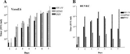
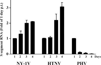
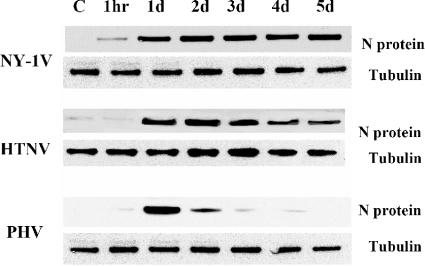
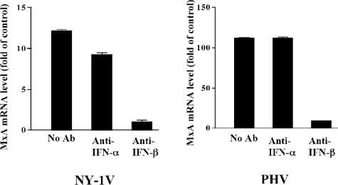
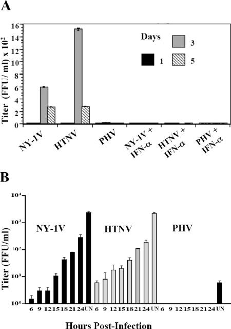
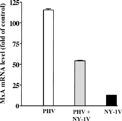
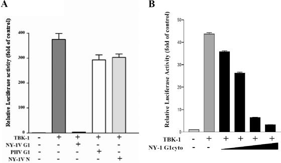
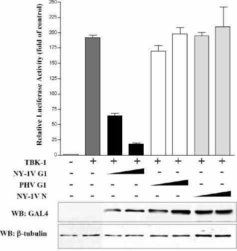
Hantaviruses cause two diseases with prominent vascular permeability defects, hemorrhagic fever with renal syndrome and hantavirus pulmonary syndrome. All hantaviruses infect human endothelial cells, although it is unclear what differentiates pathogenic from nonpathogenic hantaviruses. We observed dramatic differences in interferon-specific transcriptional responses between pathogenic and nonpathogenic hantaviruses at 1 day postinfection, suggesting that hantavirus pathogenesis may in part be determined by viral regulation of cellular interferon responses. In contrast to pathogenic NY-1 virus (NY-1V) and Hantaan virus (HTNV), nonpathogenic Prospect Hill virus (PHV) elicits early interferon responses following infection of human endothelial cells. We determined that PHV replication is blocked in human endothelial cells and that RNA and protein synthesis by PHV, but not NY-1V or HTNV, is inhibited at 2 to 4 days postinfection. The addition of antibodies to beta interferon (IFN-beta) blocked interferon-directed MxA induction by >90% and demonstrated that hantavirus infection induces the secretion of IFN-beta from endothelial cells. Coinfecting endothelial cells with NY-1V and PHV resulted in a 60% decrease in the induction of interferon-responsive MxA transcripts by PHV and further suggested the potential for NY-1V to regulate early IFN responses. Expression of the NY-1V G1 cytoplasmic tail inhibited by >90% RIG-I- and downstream TBK-1-directed transcription from interferon-stimulated response elements or beta-interferon promoters in a dose-dependent manner. In contrast, expression of the NY-1V nucleocapsid or PHV G1 tail had no effect on RIG-I- or TBK-1-directed transcriptional responses. Further, neither the NY-1V nor PHV G1 tails inhibited transcriptional responses directed by a constitutively active form of interferon regulatory factor 3 (IRF-3 5D), and IRF-3 is a direct target of TBK-1 phosphorylation. These findings indicate that the pathogenic NY-1V G1 protein regulates cellular IFN responses upstream of IRF-3 phosphorylation at the level of the TBK-1 complex. These findings further suggest that the G1 cytoplasmic tail contains a virulence element which determines the ability of hantaviruses to bypass innate cellular immune responses and delineates a mechanism for pathogenic hantaviruses to successfully replicate within human endothelial cells.


Figures

FIG. 1.
FIG. 1.
Hantavirus replication in HUVECs and Vero E6 cells. Vero E6 cells (A) and HUVECs (B) were infected with NY-1V, HTNV, or PHV at an MOI of 1. Viral titers in the supernatant of infected cells were analyzed 1 to 7 days postinfection by an infectious focus assay (16). Days postinfection are indicated on the x axis, and titers are represented as FFU/ml.
FIG. 2.
FIG. 2.
Kinetics of S segment RNA synthesis during hantavirus infection. HUVECs were mock infected or infected with NY-1V, HTNV, or PHV (MOI of 1). S segment RNA levels were determined by quantitative real-time PCR using hantavirus S segment-specific primers 1 to 4 days postinfection, and responses from duplicates were normalized to GAPDH mRNA levels. Experiments were performed twice with similar results, and mRNA levels are expressed as the change (n-fold) from 1-day levels.
FIG. 3.
FIG. 3.
Western blot analysis of N protein expression. HUVECs were infected with NY-1V, HTNV, or PHV (MOI of 1) or mock infected (C). Cells were lysed at 1 hour or 1 to 5 days postinfection as indicated. Total protein levels were determined, and an equivalent amount of whole-cell lysate was separated by 10% SDS-polyacrylamide gel electrophoresis. Proteins were detected by Western blotting using anti-nucleocapsid polyclonal rabbit antibody or anti-tubulin monoclonal antibody (Sigma), species-specific secondary antibodies (HRP conjugated), and enhanced chemiluminescence (Amersham).
FIG. 4.
FIG. 4.
Kinetics of MxA and ISG56 mRNA induction during hantavirus infection. HUVECs were infected with NY-1V or PHV (MOI of 1) or mock infected. One day postinfection, ISG56 (A) and MxA (B) mRNA levels were determined, relative to those of mock-infected controls, using quantitative real-time PCR and normalized to GAPDH mRNA levels. MxA mRNA levels were quantified 0, 1, 2, and 3 days postinfection.
FIG. 5.
FIG. 5.
Antibody to IFN-β inhibits hantavirus-directed MxA induction. HUVECs were infected at an MOI of 1 with NY-1V or PHV or mock infected. Following adsorption, anti-IFN-α or anti-IFN-β neutralizing antibodies were added to the media as indicated and MxA mRNA levels were quantified by real-time PCR as described above. MxA levels are shown as the increase (n-fold) compared to those of mock-infected controls. The scales of the y axes for NY-1V and PHV differ 10-fold. Ab, antibody.
FIG. 6.
FIG. 6.
Hantavirus replication in the presence of type I interferon. (A) Vero E6 cells were pretreated in duplicate with 1,000 IU/ml IFN-α or left untreated for 24 h prior to infection with NY-1V, HTNV, or PHV (MOI of 1) or mock infection. Hantavirus titers were determined using a focus assay 0, 1, 3, and 5 days postinfection as previously described (16). (B) HUVECs were infected as for panel A prior to the addition of 1,000 IU/ml of IFN-α 6 to 24 h postinfection or were left untreated (UN) as indicated. Three days postinfection, cell supernatants were titered on Vero E6 cells by using a focus assay (16).
FIG. 7.
FIG. 7.
NY-1V coinfection attenuates PHV-directed MxA induction. HUVECs were mock infected, infected with NY-1V or PHV (MOI of 0.5), or infected with NY-1V and PHV (MOI of 0.5 each) in duplicate. One day postinfection, MxA mRNA levels were quantified by real-time PCR and standardized to GAPDH mRNA levels as described above, and they are reported as the increase (n-fold) in mRNA levels compared to those of mock-infected controls.
FIG. 8.
FIG. 8.
NY-1V G1 cytoplasmic tail inhibits RIG-I-directed ISRE activation. (A) HEK 293 cells were transfected with an ISRE-driven luciferase reporter construct with or without cotransfection of the (N)RIG-I expression plasmid (500 ng). Cells were cotransfected with plasmids expressing the NY-1V or PHV G1 cytoplasmic tail or the NY-1V N protein (2 μg). (B) Cells were cotransfected with the (N)RIG-I expression vector (250 ng) and increasing amounts of plasmid expressing the NY-1V G1 cytoplasmic tail (0.5, 1, or 2 μg) or the control empty vector in order to transfect cells with constant amounts of total DNA. Two days posttransfection, cells were lysed, luciferase activity was assayed, and the increase (n-fold) in luciferase activity levels compared to those of controls which were not transfected with (N)RIG-I was reported after being normalized to Renilla luciferase levels. (C) Amino acid alignment of 142-residue G1 tail sequences from NY-1V (G1, positions 510 to 652) and PHV (G1, positions 513 to 655). PHV residues which differ from NY-1V are highlighted and bolded.
FIG. 9.
FIG. 9.
Hantavirus G1 cytoplasmic tail disrupts IFN-β promoter activation. (A) Duplicate wells of HEK 293 cells were transfected with an IFN-β promoter-driven luciferase reporter with or without a TBK-1 expression vector (500 ng). Cells were cotransfected with equal amounts of NY-1V or PHV G1 cytoplasmic (cyto) tail or NY-1V N protein expression vector (4 μg) as indicated. Luciferase activity was determined 48 h posttransfection and normalized to Renilla luciferase activity. Luciferase activity is reported as the increase (n-fold) compared to that of controls not transfected with TBK-1. (B) Cells were transfected with the TBK-1 expression vector as described above (250 ng), along with increasing amounts of plasmid expressing the NY-1V G1 cytoplasmic tail (0.5, 1, 2, or 4 μg) or the control empty vector in order to transfect cells with a constant amount of DNA.
FIG. 10.
FIG. 10.
NY-1V G1 cytoplasmic tail blocks TBK-1-directed ISRE activation. ISRE luciferase reporters were transfected into HEK 293 cells with or without TBK-1 expression plasmids (500 ng). Cells were cotransfected with increasing amounts of NY-1V G1 cytoplasmic tail, PHV G1 cytoplasmic tail, or NY-1V N protein expression vector (1 or 2 μg) or empty vector in order to transfect cells with a constant amount of DNA. Luciferase reporter activity was assayed as for Fig. 8 48 h posttransfection and normalized to Renilla luciferase activity, and it is reported as the increase (n-fold) compared to that of controls lacking TBK-1 activation. Protein expression levels of cells were similarly evaluated by Western blotting (WB) 48 h p.i. from parallel, comparably transfected monolayers using an anti-GAL4 antibody, and β-tubulin levels were analyzed as internal controls by Western blotting.
FIG. 11.
FIG. 11.
NY-1V G1 tail inhibits transcription upstream of IRF-3 phosphorylation. ISRE luciferase reporters were transfected into HEK 293 cells with or without IRF-3 5D expression plasmids (500 ng). Cells were cotransfected with the NY-1V or PHV G1 cytoplasmic tail or NY-1V N protein expression vector as indicated (2 μg). Luciferase reporter activity was assayed as for Fig. 8 48 h posttransfection and is reported as the increase (n-fold) compared to that of controls lacking IRF-3 5D.

References

    1. Aebi, M., J. Fah, N. Hurt, C. E. Samuel, D. Thomis, L. Bazzigher, J. Pavlovic, O. Haller, and P. Staeheli. 1989. cDNA structures and regulation of two interferon-induced human Mx proteins. Mol. Cell. Biol. 9:5062-5072. - PMC - PubMed
    1. Bai, J., K. Zhu, and G. Zhou. 1997. [The therapeutic effect of purified human leucocytic interferon-alpha on hemorrhagic fever with renal syndrome]. Zhonghua Nei Ke Za Zhi 36:90-93. (In Chinese.) - PubMed
    1. Basler, C. F., and A. Garcia-Sastre. 2002. Viruses and the type I interferon antiviral system: induction and evasion. Int. Rev. Immunol. 21:305-337. - PubMed
    1. Bridgen, A., F. Weber, J. K. Fazakerley, and R. M. Elliott. 2001. Bunyamwera bunyavirus nonstructural protein NSs is a nonessential gene product that contributes to viral pathogenesis. Proc. Natl. Acad. Sci. USA 98:664-669. - PMC - PubMed
    1. Conzelmann, K.-K. 2005. Transcriptional activation of alpha/beta interferon genes: interference by nonsegmented negative-strand RNA viruses. J. Virol. 79:5241-5248. - PMC - PubMed

Publication types

MeSH terms

LinkOut - more resources