. 2006 Oct;80(19):9730-40.
doi: 10.1128/JVI.00246-06.

ORF18 is a transfactor that is essential for late gene transcription of a gammaherpesvirus

Affiliations

ORF18 is a transfactor that is essential for late gene transcription of a gammaherpesvirus

Vaithilingaraja Arumugaswami et al. J Virol. 2006 Oct.

Abstract

Lytic replication of the tumor-associated human gammaherpesviruses Epstein-Barr virus and Kaposi's sarcoma-associated herpesvirus has important implications in pathogenesis and tumorigenesis. Herpesvirus lytic genes have been temporally classified as exhibiting immediate-early (IE), early, and late expression kinetics. Though the regulation of IE and early gene expression has been studied extensively, very little is known regarding the regulation of late gene expression. Late genes, which primarily encode virion structural proteins, require viral DNA replication for their expression. We have identified a murine gammaherpesvirus 68 (MHV-68) early lytic gene, ORF18, essential for viral replication. ORF18 is conserved in both beta- and gammaherpesviruses. By generating an MHV-68 ORF18-null virus, we characterized the stage of the virus lytic cascade that requires the function of ORF18. Gene expression profiling and quantitation of viral DNA synthesis of the ORF18-null virus revealed that the expression of early genes and viral DNA replication were not affected; however, the transcription of late genes was abolished. Hence, we have identified a gammaherpesvirus-encoded factor essential for the expression of late genes independently of viral DNA synthesis.


Figures

FIG. 1.
FIG. 1.
Analysis of recombinant viruses. (A) The location of ORF18 in the MHV-68 BAC (wt) genome is indicated. ORF18 and the flanking ORFs are shown with EcoRI (e) and AvrII (a) restriction sites. The 40-bp internal repeat sequence present in the M6 gene is shown as a striped box. (B) The position of stop codons and the AvrII insertion site in ORF18-null virus (18S) is depicted. The solid bar indicates the location of the probe used for Southern blotting. The EcoRI or AvrII restriction pattern of ORF18 locus is indicated. For AvrII, the fragments (sizes are in kilobase pairs) generated by 18S (below the line) and wt (above the line) viruses are shown. (C) Restriction profile of recombinant viruses. Five micrograms of viral BAC plasmids (two clones for each of 18R and 18S viruses) was digested with EcoRI or AvrII and separated on a 0.7% gel, and the DNA was visualized with UV after ethidium bromide staining. (D) Southern blotting of the EcoRI- or AvrII-digested viral BAC DNA. M, 1-kbp DNA ladder; TR, terminal repeat.
FIG. 2.
FIG. 2.
Rescue of ORF18-null virus by ORF18 trans-complementation. ORF18-null BAC plasmid (18S) was cotransfected into BHK-21 cells with (+) or without (−) complementing FLAG-tagged ORF18 expression vector. Wild-type MHV-68 (wt) and ORF18 revertant (18R) BAC plasmids were also transfected as positive controls. (Upper panel) At 5 days posttransfection, viral lytic antigen expression was analyzed by Western blotting using rabbit polyclonal anti-MHV-68 antisera. (Middle panel) Immunoblot probed with mouse monoclonal anti-FLAG antibody. The lower panel depicts a cellular cross-reacting antigen as a control. M, marker; UI, uninfected.
FIG. 3.
FIG. 3.
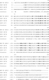
Alignment of the predicted amino acid sequences of ORF18 homologues in murine gammaherpesvirus 68 (MHV-68), Kaposi's sarcoma-associated herpesvirus (KSHV), bovine herpesvirus 4 (BHV4), and human cytomegalovirus (HCMV). The consensus sequence of the alignment is shown. Identical amino acid residues conserved among four viruses are depicted in boldface.
FIG. 4.
FIG. 4.
Mapping ORF18 regions essential for trans-complementation. (A) Schematic representation of FLAG-tagged (▪) ORF18 deletion mutants. The deleted ORF18 region is depicted as a dotted line. The N-terminal (gray boxes) and C-terminal (horizontally striped boxes) nonconserved regions as well as basic (black boxes with white dots) and hydrophobic (diagonally striped boxes) regions are shown. (B) The pFLAG-CMV2-based ORF18 deletion mutant plasmids were tested for their ability to trans-complement ORF18-null virus (18S) in BHK-21 cells. At 5 days posttransfection, the cells were subjected to Western blotting to detect the expression of viral capsid antigen ORF65 using rabbit polyclonal antibody. The ORF18 mutant proteins were detected using monoclonal anti-FLAG antibody. A nonspecific band indicated by the star serves as a loading control.
FIG. 5.
FIG. 5.
Growth kinetics of ORF18-null virus. For both single-step (A) and multiple-step (B) growth curves, noncomplementing BHK-21 cells were infected with wt, 18R, or 18S virus at MOIs of 1 and 0.01, respectively. Infected cells were harvested at the indicated time points postinfection and the viral titer, in 50% tissue culture infectivity doses (TCID50), was determined in a Tet-inducible ORF18 cell line by limiting dilution.
FIG. 6.
FIG. 6.
Viral lytic protein expression. (Upper panel) ORF18-complementing 293FT-18 and noncomplementing BHK-21 cell lines were infected with wt, 18R, or 18S virus at an MOI of 1 with (+) or without (−) PAA treatment. At 24 hpi, the infected cell lysate was harvested and the expression of viral capsid antigen ORF65 was determined by Western blotting. (Middle panel) MHV-68 lytic antigen was detected by reprobing the blot with rabbit polyclonal antisera. The lower panel depicts a cellular cross-reacting antigen as a control. M, marker; UI, uninfected.
FIG. 7.
FIG. 7.
Analysis of gene expression pattern. The BHK-21 cells were infected with wt or 18S virus. The total RNA harvested at 24 hpi was subjected to Northern blot analysis. The blot was probed with ORF57 (A), M3 (B), ORF65 (C), or cellular GAPDH (D). Note that the ORF65 mRNA is completely absent in 18S virus. Arrow heads (◂) indicate major lytic transcripts identified by each probe. The sizes of the DNA ladder are indicated on the left. M, marker.
FIG. 8.
FIG. 8.
Analysis of transcript profile. BHK-21 cells were infected with wt or 18S virus (MOI, 5). Total RNA harvested at 12 and 24 hpi was reverse transcribed, and the resulting cDNAs were hybridized to a DNA array spotted with MHV-68 genes. Array values were normalized against cellular GAPDH expression. The transcript profiles of wt and 18S viruses were compared, and the fold reduction of 18S virus gene expression was plotted for each gene on the array for 12 h and 24 h.
FIG. 9.
FIG. 9.
Viral promoter-reporter assay. The viral promoters of (A) ORF57 (early), (B) M3 (early-late), (C) ORF26 (true-late), or (D) ORF65 (true-late) driving a firefly luciferase reporter construct were individually transfected into BHK-21 cells, and at 24 h posttransfection the cells were infected with wt or 18S virus in the presence or absence of PAA. At 24 hpi, cells were lysed and luciferase activity was measured. Values were normalized against a Renilla luciferase internal control. The normalized firefly luciferase activities of viral infected cells were compared to that of uninfected cells for calculating the fold induction.
FIG. 10.
FIG. 10.
Quantitation of viral genome replication. The ORF18-complementing 293FT-18 (A and B) and noncomplementing BHK-21 cell lines (C and D) were infected with wt, 18R, or 18S virus at an MOI of 1 with or without the viral DNA polymerase inhibitor PAA. Total infected cell DNA was harvested at 12 (A and C) and 24 hpi (B and D), and the viral genome copy number per 100 ng of DNA was determined by quantitative PCR.
FIG. 11.
FIG. 11.
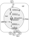
Schematic model diagram showing the stage at which ORF18 functions during gammaherpesvirus replication. The circularized virus genome in the nucleus (nuc) is shown. The expression cascade of immediate-early (IE), early (E), and late (L) transcripts are indicated with squiggly lines. ORF18 regulates the expression of late genes downstream of viral DNA synthesis. PAA is an inhibitor of virus DNA replication. cyt, cytoplasm.

References

    1. Adler, H., M. Messerle, M. Wagner, and U. H. Koszinowski. 2000. Cloning and mutagenesis of the murine gammaherpesvirus 68 genome as an infectious bacterial artificial chromosome. J. Virol. 74:6964-6974. - PMC - PubMed
    1. Ahn, J. W., K. L. Powell, P. Kellam, and D. G. Alber. 2002. Gammaherpesvirus lytic gene expression as characterized by DNA array. J. Virol. 76:6244-6256. - PMC - PubMed
    1. Amon, W., U. K. Binne, H. Bryant, P. J. Jenkins, C. E. Karstegl, and P. J. Farrell. 2004. Lytic cycle gene regulation of Epstein-Barr virus. J. Virol. 78:13460-13469. - PMC - PubMed
    1. AuCoin, D. P., K. S. Colletti, S. A. Cei, I. Papouskova, M. Tarrant, and G. S. Pari. 2004. Amplification of the Kaposi's sarcoma-associated herpesvirus/human herpesvirus 8 lytic origin of DNA replication is dependent upon a cis-acting AT-rich region and an ORF50 response element and the trans-acting factors ORF50 (K-Rta) and K8 (K-bZIP). Virology 318:542-555. - PubMed
    1. Bechtel, J. T., R. C. Winant, and D. Ganem. 2005. Host and viral proteins in the virion of Kaposi's sarcoma-associated herpesvirus. J. Virol. 79:4952-4964. - PMC - PubMed

Publication types

MeSH terms

LinkOut - more resources