. 2006 Oct;80(19):9811-21.
doi: 10.1128/JVI.00872-06.

Structural requirements for gp80 independence of human herpesvirus 8 interleukin-6 (vIL-6) and evidence for gp80 stabilization of gp130 signaling complexes induced by vIL-6

Affiliations

Structural requirements for gp80 independence of human herpesvirus 8 interleukin-6 (vIL-6) and evidence for gp80 stabilization of gp130 signaling complexes induced by vIL-6

Daming Chen et al. J Virol. 2006 Oct.

Abstract

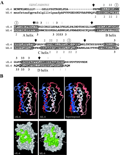
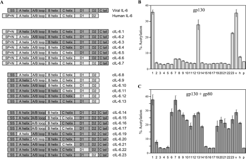
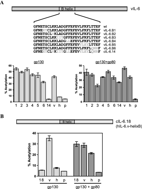
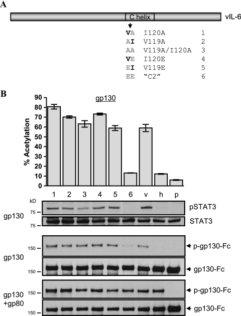
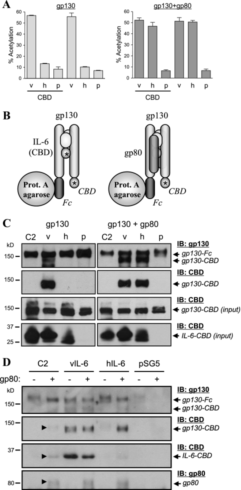
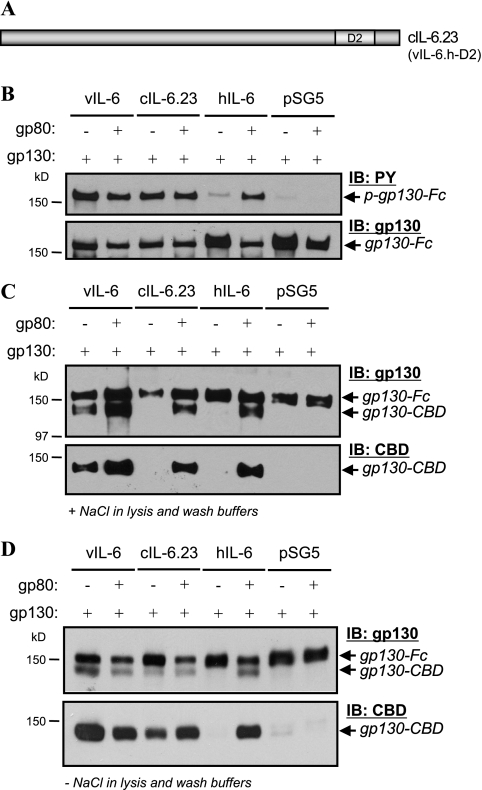
Human herpesvirus 8 interleukin-6 (vIL-6) displays 25% amino acid identity with human IL-6 (hIL-6) and shares an overall four-helix-bundle structure and gp130-mediated STAT/mitogen-activated protein kinase signaling with its cellular counterpart. However, vIL-6 is distinct in that it can signal through gp130 alone, in the absence of the nonsignaling gp80 alpha-subunit of the IL-6 receptor. To investigate the structural requirements for gp80 independence of vIL-6, a series of expression vectors encoding vIL-6/hIL-6 chimeric and site-mutated IL-6 proteins was generated. The replacement of hIL-6 residues with three vIL-6-specific tryptophans implicated in gp80 independence from crystallographic studies or the A and C helices containing these residues did not confer gp80 independence to hIL-6. The N- and C-terminal regions of vIL-6 could be substituted with hIL-6 sequences with the retention of gp80-independent signaling, but substitutions of other regions of vIL-6 (helix A, A/B loop, helix B, helix C, and proximal half of helix D) with equivalent sequences of hIL-6 abolished gp80 independence. Interestingly, the B helix of vIL-6 was absolutely required for gp80 independence, despite the fact that this region contains no receptor-binding residues. Point mutational analysis of helix C, which contains residues involved in physical and functional interactions with gp130 domains 2 and 3 (cytokine-binding homology region), identified a variant, VI120EE, that was able to signal and dimerize gp130 only in the presence of gp80. gp80 was also found to stabilize gp130:g130 dimers induced by a distal D helix variant of vIL-6 that was nonetheless able to signal independently of gp80. Together, our data reveal the crucial importance of overall vIL-6 structure and conformation for gp80-independent signaling and provide functional and physical evidence of the stabilization of vIL-6-induced gp130 signaling complexes by gp80.


Figures

FIG. 1.
FIG. 1.
Primary and three-dimensional structures of viral and human IL-6. (A) Alignment of the amino acid sequences specified by the human and HHV-8 IL-6 ORFs. Indicated are the α-helices, residues predicted (vIL-6 site I) or demonstrated to interact with gp80 (“1”) and gp130 (“2” and “3”), and vIL-6-specific site II tryptophan residues (circled) predicted to contribute significantly to site II interactions with gp130 CHR (domains 2 and 3) (4, 5, 11). Site III residues of IL-6 interact with gp130 Ig-like domain 1, and site I residues with CHR residues of gp80. Also shown (arrows) are the junctions of splices between vIL-6 and hIL-6 sequences for the generation of IL-6 chimeric proteins (listed in Fig. 3A). The cleaved signal sequence of hIL-6 is indicated in lowercase. (B) Three-dimensional structures of vIL-6 (5) and hIL-6 (11) as illustrated by ribbon and “skin” models, orientated with helices A and C in the front plane to show the site II interface. The ribbon diagram on the right shows the vIL-6 and hIL-6 structures superimposed to emphasize the highly conserved secondary and tertiary structures of the two cytokines. The skin models below are orientated equivalently to the ribbon diagrams and show site II residues (shaded) of vIL-6 and hIL-6, including the three vIL-6-specific tryptophan residues and colinear residues of hIL-6 (labeled). Numbering of hIL-6 residues is from the first amino acid of the mature, cleaved protein.
FIG. 2.
FIG. 2.
Functional analyses of hIL-6 proteins with introduced vIL-6-specific site II tryptophan residues and site II-containing vIL-6 helices A and C and vIL-6 proteins with the tryptophans altered to alanine residues. (A) The altered vIL-6 and hIL-6 proteins that were generated are indicated. Mutated residues correspond to W41, W44, and W134 of vIL-6 and Y32, D35, and Q125 of hIL-6. Each point-mutated or chimeric ORF was cloned into pSG5 to allow the expression of the encoded proteins in transfected eukaryotic cells. (B) Data from reporter-based assays utilizing the STAT-responsive reporter pα2MCAT (24) and expression vectors for gp80 and gp130. These vectors were cotransfected into HEK293T cells with each of the IL-6 expression constructions listed in panel A (1 to 4; v, vIL-6; h, hIL-6) or pSG5 (p, negative control) to determine the signaling activities of the IL-6 proteins via gp130 or gp130+gp80, as described previously (26). The mutation of the site II tryptophans in vIL-6 reduced but did not eliminate gp80-independent signaling by the viral cytokine, and substitution of the A and C helices of hIL-6 with those of vIL-6 had no effect on gp80 dependence of hIL-6. Introduction of the three site II tryptophans into hIL-6 allowed modest gp80-independent signaling by hIL-6. Transfections were carried out in triplicate. Error bars indicate standard deviations.
FIG. 3.
FIG. 3.
Generation and analyses of vIL-6/hIL-6 chimeras to identify regions involved in gp80-independent signaling by vIL-6. (A) Diagrammatic illustration of vIL-6/hIL-6 chimeric ORFs generated and cloned into pSG5 for eukaryotic expression. SS, signal sequence; SP+N, signal peptide plus N terminus. (B) Functional analyses of chimeric proteins relative to native vIL-6 (v) and hIL-6 (h) by STAT-CAT reporter (pα2M-CAT) assays to detect signaling through overexpressed gp130. Numbers correspond to designations given to each of the chimeric constructions in panel A. Cultures transfected with pSG5 (p) were used as negative controls. Error bars indicate standard deviations. (C) An experiment similar to that described for panel B was carried out to assay for signaling activities of each of the chimeric proteins through overexpressed gp130+gp80. Triplicate transfections were carried out for each chimera in each experiment. Error bars indicate standard deviations.
FIG. 4.
FIG. 4.
Contribution of vIL-6 helix B to gp80-independent signaling. (A) Generation and analysis of B helix substitution variants of vIL-6. Single or grouped codon substitutions were introduced into vIL-6 to match the equivalent colinear codons in hIL-6. These altered ORFs were then cloned into pSG5. Reporter assays were carried out to determine the effects of the introduced B helix changes on gp80-independent signaling by vIL-6. Assays for signaling in gp80+gp130-overexpressing cells were performed in parallel. None of the introduced paired substitutions significantly altered gp80-independent signaling by vIL-6. (B) Coding sequences for the B helix of hIL-6 were replaced by those of vIL-6, and the resulting encoded protein was tested for its ability to signal via gp130 alone or via gp130+gp80. The B helix of vIL-6 could not confer gp80 independence to hIL-6. All transfections were performed in triplicate. Error bars indicate standard deviations. v, vIL-6; h, hIL-6; p, pSG5.
FIG. 5.
FIG. 5.
Mutational analysis of the C helix of vIL-6. (A) Pairwise substitution mutagenesis of the C helix of vIL-6. Substitutions introduced into the vIL-6 ORF match colinear hIL-6 codons. (B) Positions within the three-dimensional structure of vIL-6 of C helix site II residues involved in interactions with gp130-CHR. (C) gp130 and gp130+gp80 reporter-based signaling assays to determine the effects of the introduced amino acid substitutions on gp80-independent signaling by vIL-6. All but the “C2” variant (VI120EE) were able to signal at around wild-type levels through overexpressed gp130 in the absence of overexpressed gp80. Error bars indicate standard deviations. (D) Parallel Western blot-based assays to identify directly vIL-6-induced STAT3 activation in cells overexpressing gp130 or gp130+gp80. Cells were cotransfected with receptor and vIL-6 expression constructions and harvested 48 h posttransfection. Tyrosine-phosphorylated (active) STAT3 (“pSTAT3”) was detected by immunoprobing using a phosphospecific antibody; total STAT3 was detected on stripped blots using a phosphorylation-independent antibody to STAT3. (E) Western analysis for g130 phosphorylation in cells cotransfected with ligand and receptor expression constructions. Here, a gp130-Fc expression vector was used to allow direct protein A-agarose precipitation of gp130 from cell extracts. A phosphotyrosine (PY) antibody was used to detect phosphorylated gp130, and total precipitated gp130-Fc was detected on stripped blots using an appropriate antibody to gp130.
FIG. 6.
FIG. 6.
Signaling by C2-position point variants of vIL-6. (A) Further mutations at positions 119 and 120 were generated as indicated. (B) These variants were tested in pα2MCAT reporter and phospho-STAT3 Western blot assays for their abilities to signal via gp130 independently of gp80. Reporter assay transfections were performed in triplicate. Error bars indicate standard deviations. v, vIL-6; h, hIL-6; p, pSG5.
FIG. 7.
FIG. 7.
Dimerization of gp130 by vIL-6.C2. (A) Reporter (pα2MCAT) assays were undertaken, as outlined for previous experiments, to confirm the functional integrities of vIL-6-CBD (v) and hIL-6-CBD (h) fusion proteins used in the gp130 dimerization assays. Empty expression plasmids (p) pSG5-CBD and pSG5 were used as negative controls for the CBD fusion and native proteins, respectively. Values shown are averages from duplicate transfections, with bars indicating the range. (B) Diagrammatic representation of coprecipitation experiments performed to analyze gp130 dimerization induced by vIL-6.C2. HEK293T cells were cotransfected with expression vectors for gp130-Fc, gp130-CBD, and IL-6-CBD (vIL-6, vIL-6.C2, or hIL-6), or pSG5-CBD (negative control), with or without gp80 expression vector. Protein (prot.) A-agarose was used to precipitate gp130-Fc from cell lysates, and this and coprecipitated gp130-CBD (indicative of gp130 dimerization) were identified by Western blotting using antibodies specific for gp130 or CBD. (C) Assays for gp130 dimerization induced by vIL-6.C2 relative to wild-type vIL-6 and hIL-6. Stable, gp130-Fc-containing complexes were precipitated directly from cell lysates, and PBS was used to wash protein A-agarose-bound complexes prior to gel loading. (D) Cells were treated with protein cross-linker (DSP, 0.1 mM) prior to cell lysis and protein A-agarose precipitation of gp130-Fc-containing complexes. Cross-linking was reversed in 5% β-mercaptoethanol before gel loading. +, presence of; −, absence of; IB, immunoblot.
FIG. 8.
FIG. 8.
Analysis of gp130 dimerization and phosphorylation induced by cIL-6.23 (vIL-6.h-D2) in the absence (+) and presence (−) of gp80. (A) Diagrammatic representation of cIL-6.23, in which the distal half of helix D of vIL-6 is replaced by that of hIL-6. (B) Verification of gp80-independent signaling by cIL-6.23 using gp130 phosphorylation assay. gp130-Fc was precipitated from HEK293T cells cotransfected with gp130-Fc vector with or without gp80 vector and pSG5-based expression plasmids for vIL-6, vIL-6.23, or hIL-6; empty vector, pSG5, was used as a negative control. Precipitated gp130-Fc was subjected to PAGE and blotted onto membrane, and phosphotyrosine (PY) residues were detected by using anti-PY antibody. (C) gp130-Fc-based coprecipitation assays for gp130 dimerization, showing gp80-dependent coprecipitation of coexpressed gp130-CBD from lysates of cells expressing vIL-6.23. Wild-type vIL-6 and hIL-6 were used as positive controls for gp80-independent and gp80-dependent gp130 dimerization, respectively, and empty pSG5 vector provided the negative control. Cells were lysed in PBS containing 0.1% NP-40, and precipitates were washed with PBS prior to gel loading. (D) Repeat of the experiment described for panel C except that salt-free TE buffer was used to lyse cells and wash protein A-bound complexes prior to gel loading. IB, immunoblot.

References

    1. Aoki, Y., E. S. Jaffe, Y. Chang, K. Jones, J. Teruya-Feldstein, P. S. Moore, and G. Tosato. 1999. Angiogenesis and hematopoiesis induced by Kaposi's sarcoma-associated herpesvirus-encoded interleukin-6. Blood 93:4034-4043. - PubMed
    1. Aoki, Y., M. Narazaki, T. Kishimoto, and G. Tosato. 2001. Receptor engagement by viral interleukin-6 encoded by Kaposi's sarcoma-associated herpesvirus. Blood 98:3042-3049. - PubMed
    1. Aoki, Y., R. Yarchoan, K. Wyvill, S. Okamoto, R. F. Little, and G. Tosato. 2001. Detection of viral interleukin-6 in Kaposi's sarcoma-associated herpesvirus-linked disorders. Blood 97:2173-2176. - PubMed
    1. Boulanger, M. J., D. C. Chow, E. E., Brevnova, and K. C. Garcia. 2003. Hexameric structure and assembly of the interleukin-6/IL-6α-receptor/gp130 complex. Science 300:2101-2104. - PubMed
    1. Boulanger, M. J., D. C. Chow, E. Brevnova, M. Martick, G. Sandford, J. Nicholas, and K. C. Garcia. 2004. Molecular mechanisms for viral mimicry of a human cytokine: activation of gp130 by HHV-8 interleukin-6. J. Mol. Biol. 335:641-654. - PubMed

Publication types

MeSH terms