

Optical coherence tomography of the anterior segment in secondary glaucoma with corneal opacity after penetrating keratoplasty
- PMID: 16973665
- PMCID: PMC1857632
- DOI: 10.1136/bjo.2006.100099
Optical coherence tomography of the anterior segment in secondary glaucoma with corneal opacity after penetrating keratoplasty
Abstract
Aim: To evaluate secondary glaucoma after penetrating keratoplasty with anterior-segment optical coherence tomography (OCT).
Design: Case series.
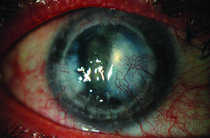
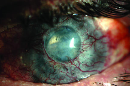
Methods: Four eyes of four patients with corneal opacity and increased intraocular pressure (IOP) were evaluated using high-speed (2000 axial scans/s) OCT at 1.3 microm wavelength. Cross-sectional images of the anterior segment were analysed to assess the cause of increase in pressure.
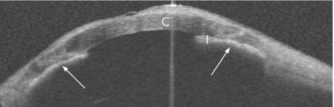
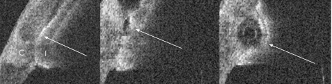
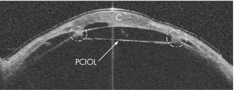
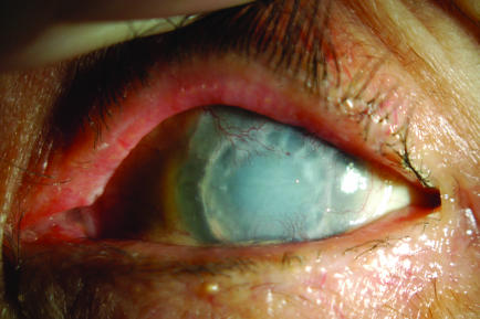
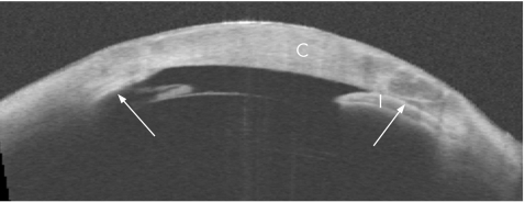
Results: Slit-lamp evaluation of the anterior chamber in all cases was limited by corneal opacity. The OCT imaging allowed visualisation of anterior-segment structures behind the opaque corneas. Using OCT, iris-intraocular lens adhesion and pupillary block were identified as the probable reasons for the increased IOP in one case. Peripheral anterior synechiae and angle closure were identified in the three remaining cases. In two cases, we found that the tip of the aqueous drainage tube was blocked by peripheral anterior synechiae.
Conclusions: OCT is similar to ultrasound in that it allows visualisation through opaque corneas. However, OCT has an advantage in that it requires neither contact nor immersion. It is a valuable tool for evaluating the depth of the anterior chamber angle and the causes of secondary angle closure.
Conflict of interest statement
Competing interests: DH receives royalty from OCT patents licensed to Carl Zeiss Meditec, Dublin, California, USA. D H and Y L receive research grant support from Carl Zeiss Meditec.
References
-
- Irvine A R, Kaufman H E. Intraocular pressure following penetrating keratoplasty. Am J Ophthalmol 196968835–844. - PubMed
-
- Lee R K, Fantes F. Surgical management of patients with combined glaucoma and corneal transplant surgery. Curr Opin Ophthalmol 20031495–99. - PubMed
-
- Krontz D P, Wood T O. Corneal decompensation following acute angle‐closure glaucoma. Ophthalmic Surg 198819334–338. - PubMed
Publication types
MeSH terms
Grants and funding
LinkOut - more resources
Full Text Sources