. 2006 Sep 26;103(39):14343-8.
doi: 10.1073/pnas.0606480103. Epub 2006 Sep 14.

The crystal structure of human IRE1 luminal domain reveals a conserved dimerization interface required for activation of the unfolded protein response

Affiliations

The crystal structure of human IRE1 luminal domain reveals a conserved dimerization interface required for activation of the unfolded protein response

Jiahai Zhou et al. Proc Natl Acad Sci U S A. .

Abstract

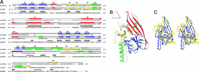
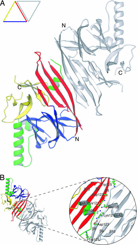
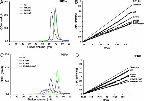
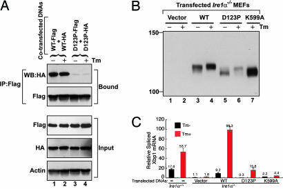
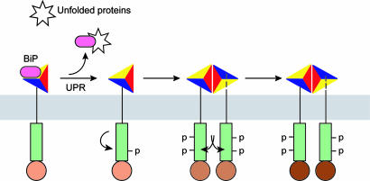
The unfolded protein response (UPR) is an evolutionarily conserved mechanism by which all eukaryotic cells adapt to the accumulation of unfolded proteins in the endoplasmic reticulum (ER). Inositol-requiring kinase 1 (IRE1) and PKR-related ER kinase (PERK) are two type I transmembrane ER-localized protein kinase receptors that signal the UPR through a process that involves homodimerization and autophosphorylation. To elucidate the molecular basis of the ER transmembrane signaling event, we determined the x-ray crystal structure of the luminal domain of human IRE1alpha. The monomer of the luminal domain comprises a unique fold of a triangular assembly of beta-sheet clusters. Structural analysis identified an extensive dimerization interface stabilized by hydrogen bonds and hydrophobic interactions. Dimerization creates an MHC-like groove at the interface. However, because this groove is too narrow for peptide binding and the purified luminal domain forms high-affinity dimers in vitro, peptide binding to this groove is not required for dimerization. Consistent with our structural observations, mutations that disrupt the dimerization interface produced IRE1alpha molecules that failed to either dimerize or activate the UPR upon ER stress. In addition, mutations in a structurally homologous region within PERK also prevented dimerization. Our structural, biochemical, and functional studies in vivo altogether demonstrate that IRE1 and PERK have conserved a common molecular interface necessary and sufficient for dimerization and UPR signaling.


Conflict of interest statement

The authors declare no conflict of interest.

Figures

Fig. 1.
Fig. 1.
The crystal structure of human IRE1α NLD. (A) Sequence and secondary structure alignment of IRE1 and PERK. The NLD sequences of human IRE1α, S. cerevisiae Ire1p, and murine PERK were aligned by using the program T-Coffee (32). Secondary structural elements are indicated above the sequence: α-helices are drawn as rectangles, β-strands as arrows, other elements as solid lines, and structurally unobserved residues as dashed lines. These elements are colored based on their locations in the structure (see Results for details). Predicted α-helices and β-strands for PERK are indicated with Greek letters. (B) A ribbon drawing of the NLD monomer. The secondary structural elements are labeled and colored as in A: α-helices are lettered and drawn as coils, β-strands are numbered and drawn as arrows, and other elements are drawn as tubes. (C) A stereo diagram showing Cα trace superimposition of human IRE1α NLD (blue) and S. cerevisiae Ire1p NLD (yellow) structures. The programs Ribbons (33) and Grasp (34) were used to produce B and C, respectively.
Fig. 2.
Fig. 2.
The molecular dimer structure of human IRE1α NLD. (A) Ribbon drawing of the NLD dimer looking straight down the twofold axis of symmetry. One subunit is colored as in Fig. 1B, and the other is gray. In the displayed orientation both C termini of the dimer would project into the page. (B) An enlarged view of the dimer interface after rotating the dimer in A 180° around the horizontal axis. Side chains of residues examined in our mutagenesis studies are shown as ball-and-stick models. The program Ribbons (33) was used to produce both drawings.
Fig. 3.
Fig. 3.
The luminal domains of IRE1 and PERK share a similar dimerization mechanism. (A and C) Gel-filtration analysis. WT human IRE1α NLD eluted as a 158-kDa protein upon gel filtration (black line), and mutants Q105E (red line), D123P (green line), and W125A (blue line) eluted 6 ml later (A). WT murine PERK NLD eluted as a 181-kDa protein (black line), and mutants K194P (red line), L196P (blue line), and K194P/L196P (green line) eluted 10 ml later (C). (B and D) Analytical ultracentrifugation sedimentation equilibrium analysis. Point mutations in human IRE1α NLD (B) and murine PERK NLD (D) shift the dimer/monomer equilibrium toward the monomeric species. The IRE1α NLD mutant D123P and the PERK NLD double mutant K194P/L196P were exclusively monomeric.
Fig. 4.
Fig. 4.
Human IRE1α mutant D123P is defective in dimerization, autophosphorylation, and RNase activity. (A) COS-1 cells were cotransfected with plasmids expressing human IRE1α (WT or D123P) tagged with Flag and HA epitopes. Lysates were prepared from cells treated with or without Tm for 4 h and subjected to immunoprecipitation using anti-Flag monoclonal antibody and then analyzed by Western blot using anti-HA antibody. (B) Ire−/− MEF cell lines stably expressing human IRE1α WT or mutant proteins were treated with or without Tm for 5 h. Cell lysates were prepared and subjected to immunoprecipitation by using anti-IRE1α antibody and then analyzed by Western blot with antibodies against IRE1α. (C) Quantitative real-time RT-PCR analysis of spliced Xbp1 transcripts. WT and Ire1α−/− MEF cell lines stably expressing WT or mutant human IRE1α proteins were treated with Tm (10 μg/ml) for 5 h, and RNA was isolated for quantitative real-time RT-PCR by using a primer set flanking the intron in the Xbp1 mRNA. The columns and bars represent the means and standard deviations of three independent experiments.
Fig. 5.
Fig. 5.
Model for staged activation of IRE1. The model depicts that several steps mediate IRE1 activation including BiP release, initial intramolecular autophosphorylation, dimerization, and dimerization-induced trans-autophosphorylation (see Discussion for details). Increasing degrees of IRE1 autophosphorylation cause higher levels of RNase activity indicated as darker shades of brown.

Similar articles

Cited by

References

    1. Schroder M, Kaufman RJ. Annu Rev Biochem. 2005;74:739–789. - PubMed
    1. Patil C, Walter P. Curr Opin Cell Biol. 2001;13:349–355. - PubMed
    1. Harding HP, Calfon M, Urano F, Novoa I, Ron D. Annu Rev Cell Dev Biol. 2002;18:575–599. - PubMed
    1. Mori K. Traffic. 2003;4:519–528. - PubMed
    1. Cox JS, Shamu CE, Walter P. Cell. 1993;73:1197–1206. - PubMed

Publication types

MeSH terms

Associated data