. 2006;34(17):4857-65.
doi: 10.1093/nar/gkl564. Epub 2006 Sep 14.

Human replication protein A unfolds telomeric G-quadruplexes

Affiliations

Human replication protein A unfolds telomeric G-quadruplexes

Tonatiuh Romero Salas et al. Nucleic Acids Res. 2006.

Abstract

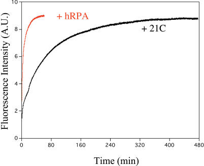
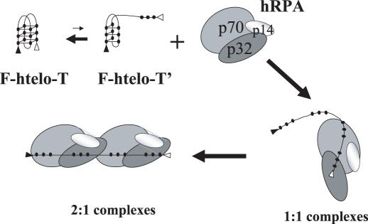
G-quadruplex structures inhibit telomerase activity and must be disrupted for telomere elongation during S phase. It has been suggested that the replication protein A (RPA) could unwind and maintain single-stranded DNA in a state amenable to the binding of telomeric components. We show here that under near-physiological in vitro conditions, human RPA is able to bind and unfold G-quadruplex structures formed from a 21mer human telomeric sequence. Analyses by native gel electrophoresis, cross-linking and fluorescence resonance energy transfer indicate the formation of both 1:1 and 2:1 complexes in which G-quadruplexes are unfolded. In addition, quadruplex opening by hRPA is much faster than observed with the complementary DNA, demonstrating that this protein efficiently unfolds G-quartets. A two-step mechanism accounting for the binding of hRPA to G-quadruplexes is proposed. These data point to the involvement of hRPA in regulation of telomere maintenance.


Figures

Figure 1
Figure 1
Schematic representation of quadruplex and duplex structures. (a) Different possible folding topologies of htelo. Closed circles depict guanines, TTA loops represent single-stranded DNA regions. (b) Schematic representation of a 21 bp duplex formed between F-htelo-T and its complementary sequence 21C where cytosines are depicted by gray circles. FLUO and TAMRA are depicted by closed and open triangles, respectively.
Figure 2
Figure 2
Titration of htelo as a function of hRPA concentration. 32P-htelo (90 nM) was incubated with various amounts of hRPA (from 45 to 630 nM) in the presence of 50 mM NaCl (a) or KCl (b) and separated on a native 5% polyacrylamide gel. CI and CII represent 1:1 and 2:1 hRPA–htelo complexes, respectively. r is the molar ratio hRPA/htelo.
Figure 3
Figure 3
Determination of the hRPA/htelo ratios in complexes. 32P-htelo (90 nM) was incubated with 90 or 630 nM of hRPA in the presence of 50 mM NaCl and separated by native 5% PAGE. Analysis using a Phosphorimager revealed radioactive htelo (a), and densitometric analysis revealed colored hRPA (b). The boxes show the areas considered to determine the hRPA/htelo ratios.
Figure 4
Figure 4
Comparison of htelo and T21 titrations by hRPA in the absence or presence of glutaraldehyde. 32P-DNA (90 nM) was incubated with various amounts of hRPA (from 90 to 630 nM) in the presence of 50 mM NaCl then cross-linked by the addition of 0.1% glutaraldehyde for 10 min. Individual reaction mixtures were analyzed on a native 5% polyacrylamide gel. (a) EMSA results after incubation of htelo. (b) EMSA results after incubation of T21. r is the molar ratio hRPA/DNA. CI and CII represent the non-covalent 1:1 and 2:1 hRPA–DNA complexes, respectively. CI, CII and CIII represent the covalent 1:1, 2:1 and putative 3:1 hRPA–DNA complexes, respectively.
Figure 5
Figure 5
Quantification of f0, f1 and f2 fractions as a function of r. The relative weights in solution of free htelo (f0, solid line), complex CI (f1, long dashed line) and complex CII (f2, short dashed line) were quantified for each r-value from hRPA–htelo EMSA (Figure 2) in the presence of Na+ (a) or K+ (b). The relative errors vary from 20% for the low r-values (r = 0.5–1) to 10% for the high r-values (r > 1). r is the molar ratio hRPA/htelo.
Figure 6
Figure 6
hRPA binding leads to G-quadruplex unfolding. Fluorimetric titration of 90 nM of F-htelo-T by increasing amounts of hRPA was performed in the presence of Na+ (a) or K+ (b). The spectra were recorded after 2 min of incubation. (c) Increasing of P as a function of the hRPA/F-htelo-T ratio (r) in the presence of Na+ (solid line) or K+ (dashed line) indicates unfolding of F-htelo-T by addition of hRPA. P was calculated from experimental FRET data: P = ID/(ID + IA), where ID and IA are the average intensities of the donor (FLUO) and acceptor (TAMRA), respectively. For each P-value, error bar is ±0.05. AU, arbitrary units.
Figure 7
Figure 7
Comparison of the F-htelo-T opening by 21C and hRPA. Each solution containing 90 nM F-htelo-T was mixed at time zero with a 5× molar excess of its complementary sequence 21C (black curve) or hRPA (red curve). Fluorescence intensity was recorded at 516 nm in the presence of 50 mM KCl. AU, arbitrary units.
Figure 8
Figure 8
Sequential model of hRPA binding to G-quadruplexes. hRPA binding is directed by single-stranded regions generated at the extremities of the partially unfolded structures as represented by F-htelo-T′. This first binding step destabilizes the G-quadruplex structure to form 1:1 complexes. This unfolded DNA bound to one hRPA molecule at the extremity facilitates binding of a second hRPA molecule to produce 2:1 complexes.

Similar articles

Cited by

References

    1. Blackburn E.H. Structure and function of telomeres. Nature. 1991;350:569–573. - PubMed
    1. Blackburn E.H. Telomere states and cell fates. Nature. 2000;408:53–56. - PubMed
    1. Li B., Oestreich S., de Lange T. Identification of human Rap1: implications for telomere evolution. Cell. 2000;101:471–483. - PubMed
    1. van Steensel B., Smogorzewska A., de Lange T. TRF2 protects human telomeres from end-to-end fusions. Cell. 1998;92:401–413. - PubMed
    1. Price C.M., Cech T.R. Telomeric DNA–protein interactions of Oxytricha macronuclear DNA. Genes Dev. 1987;1:783–793. - PubMed

Publication types