. 2006 Oct 13;99(8):861-9.
doi: 10.1161/01.RES.0000245188.41002.2c. Epub 2006 Sep 14.

Human pulmonary valve progenitor cells exhibit endothelial/mesenchymal plasticity in response to vascular endothelial growth factor-A and transforming growth factor-beta2

Affiliations

Human pulmonary valve progenitor cells exhibit endothelial/mesenchymal plasticity in response to vascular endothelial growth factor-A and transforming growth factor-beta2

Sailaja Paruchuri et al. Circ Res. .

Abstract

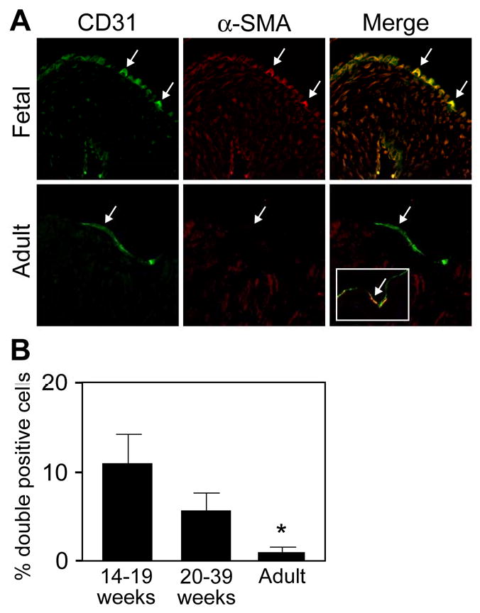
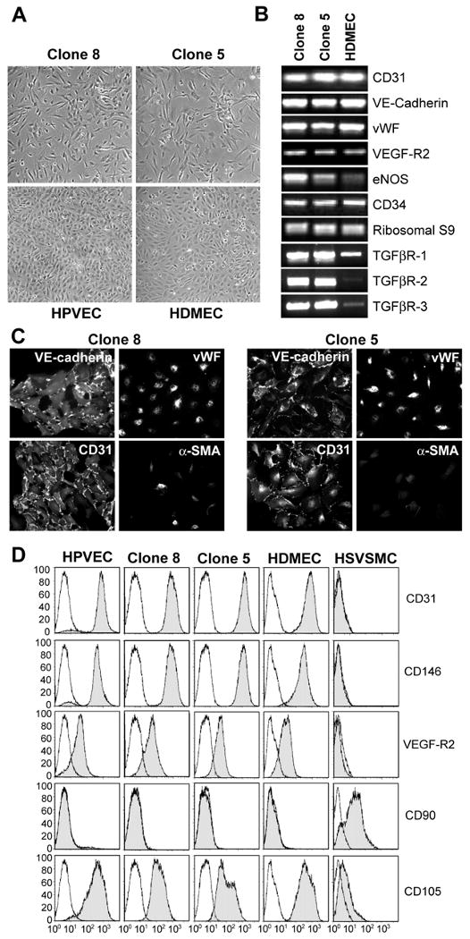
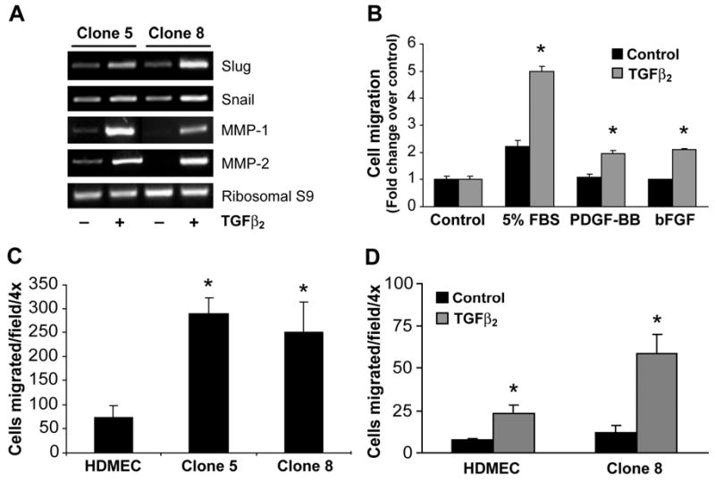
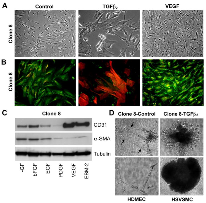
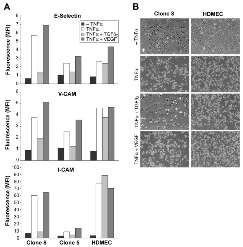
In situ analysis of fetal semilunar valve leaflets has revealed cells coexpressing endothelial and mesenchymal markers along the endothelium, with diminished frequency seen in adult valves. To determine whether such cells are progenitor cells, we isolated clonal populations from human pulmonary valves. The clones expressed endothelial markers but showed potential to further differentiate into endothelium in response to vascular endothelial growth factor (VEGF)-A. When exposed to transforming growth factor (TGF)-beta2, individual clones adopted a mesenchymal phenotype to varying degrees and expressed markers of endothelial to mesenchymal transformation (EMT). Both VEGF- and TGFbeta2-induced phenotypic changes were partially reversible, indicating the plasticity of these cells. When challenged with VEGF or TGFbeta2, a hierarchy of endothelial/mesenchymal potential could be seen among the clonal populations: cells initially closer to an endothelial phenotype showed a strong response to TGFbeta2 that could be inhibited by VEGF, whereas cells closer to a mesenchymal phenotype responded to TGFbeta2 but were resistant to endothelial-inducing effects of VEGF. These findings suggest the presence of bipotential valve progenitor cells with ability to differentiate into either endothelial or interstitial cells of the valve leaflet. Understanding the differentiation potential and function of these cells may be important for understanding heart valve disease and may also be applied to current paradigms for creating tissue-engineered heart valves.


Figures

Figure 1
Figure 1
Cells co-expressing CD31 and α-SMA in fetal and adult semilunar valves (A) Sections were double-labeled with anti-CD31 (green) and anti-α-SMA (red). Arrows indicate double-positive cells along the arterial side of the leaflet. The inset in the lower right panel shows rare double-positive cells detected in adult valves. (B) Cells co-expressing both markers were quantitated: fetal valves from 14-19 weeks gestation, 10.8 ± 3.3% (n=11); 20-39 weeks gestation, 5.6 ± 2.0%, (n=10); and adult valves, 1.0 ± 0.5%, (n=10). Data are presented as mean ± SEM. P values: 14-19 weeks versus 20-39 weeks, not significant; 14-19 weeks versus adult, p<0.05; 20-39 week versus adult, p<0.05.
Figure 2
Figure 2
Clonal cell populations from human pulmonary valve express endothelial-specific markers. (A) Phase contrast micrographs of Clone 8 and Clone 5 (top panels) parental HPVECs and HDMECs (bottom panels) grown in EBM-2. (B) RT-PCR analysis of Clones 8 (lane 1), 5 (lane 2) and HDMECs (lane 3) for endothelial-specific transcripts and TGF-β-receptor transcripts. Ribosomal S9 served as a control. (C) Immunostaining of Clone 8 (left panels) and Clone 5 (right panels) with anti-VE-cadherin, anti-vWF, anti-CD31, and anti-α-SMA Abs. (D) Flow cytometric analysis of parental HPVEC, Clone 8, Clone 5, HDMEC and HSVSMC stained with FITC-conjugated CD31, CD146, VEFG-R2, CD90 and CD105 Abs.
Figure 3
Figure 3
TGFβ2-induced expression of α-SMA and calponin. (A) Clones 8 and 5, the parental HPVEC and HDMECs were grown for 10 days in absence (control, lane 1) or presence of 2ng/ml of TGFβ1 (lane 2), TGFβ2 (lane 3) or TGFβ3 (lane 4). Cell lysates were analyzed by western blot for the expression of CD31, α-SMA and calponin. (B) Clone 8 (left panels) and Clone 5 (right panels) were grown for 10 days in absence (control; top panels) or presence of 2ng/ml of TGFβ2 (TGFβ2; bottom panels). Cells were double-labeled with goat anti-human CD31/FITC-conjugated secondary Ab and mouse anti-α-SMA/Texas Red-conjugated secondary Ab. α-SMA positive cells, which also expressed CD31, were counted to determine number of positive cells in each clone. * P values<0.05.
Figure 4
Figure 4
TGFβ2-induced expression of EMT markers, migration, and invasion. (A) Clone 5 (lanes1 and 2) and Clone 8 (lanes 3 and 4) were grown for 10 days in absence (lanes 1 and 3) or presence of 2 ng/ml TGFβ2 (lanes 2, 4). RNA was extracted and RT-PCR was performed with Slug, Snail, MMP-1 and MMP-2 primers. Ribosomal S9 served as a control. (B) Clone 8 cells cultured in absence (black bars) or presence (grey bars) of TGFβ2 for 10 days were tested for ability to migrate towards EBM (control), EBM with 5% FBS, 10 ng/ml PDGF-BB, and 10ng/ml bFGF. (C) HDMECs, Clone 5 and Clone 8 cells were cultured in EBM-2 medium and basal migration towards control medium was tested. (D) HDMECs and Clone 8 cells were cultured in absence (black bars) or presence (grey bars) of TGFβ2 in EBM-2 medium for 10 days and were tested for invasion into collagen gels for 72 h. Data from the migration and invasion assays are mean ± SD of two independent experiments performed in triplicates. In panels B-D, * represent p<0.05 compared control.
Figure 5
Figure 5
Phenotypic modulation of HPVEC clones. Clone 8 cells were cultured in absence (control) or presence of 2ng/ml TGFβ2 or 10ng/ml VEGF in EBM-2 medium for 10 days. (A) Phase contrast micrographs show cell morphology. (B) Double-label immunostaining of same cells with anti-CD31 (FITC) and anti-α-SMA (Texas Red). (C) Clone 8 cells were cultured in growth factor depleted medium (-GFl) or supplemented with bFGF (10ng/ml), EGF (10ng/ml), PDGF (10ng/ml), VEGF (10ng/ml), and EBM-2 medium for 10 days. Cell lysates were analyzed for the expression of CD31 and α-SMA, and tubulin to confirm equal loading. (D) Tube formation on Matrigel. Clone 8 cells were cultured in absence (control) or presence of TGFβ2 in EBM-2 medium for 10 days and then plated on Matrigel. Tube formation was assessed after 24 hours in comparison with HDMEC and HSVSMC that had not been treated with TGFβ2.
Figure 6
Figure 6
Cytokine-induced expression of leukocyte adhesion molecules and adherence of leukocytes. Clone 8 cells were cultured in absence or presence of 2ng/ml TGFβ2 or 10ng/ml VEGF in EBM-2 medium for 10 days. Cells were further treated without (-TNFα) or with TNF-α (TNF-α +) for 5 h and analyzed for the expression of leukocyte adhesion molecules by flow cytometry. B. HL-60 cell adhesion to Clone 8 and HDMECs with similar treatments as in A was determined by light microscope.
Figure 7
Figure 7
Changes induced by TGFβ2 and VEGF were reversible and effects of ECM on TGFβ2-induced EMT. Panel A: Schematic of culture conditions: Clone 8 cells were cultured in EBM-2 medium (C), EBM-2 medium with 2ng/ml TGFβ2 (T) or EMB-2 medium with 10ng/ml VEGF (V) for 10 days. Cells were then re-plated and cultured for 10 days in (C), (T) or (V). Panel B: Cell lysates were analyzed for the expression of CD31 and α-SMA. It is important to note that the EBM-2 medium (C) is supplemented with the commercially available SingleQuots, which contains 2-5 ng/ml VEGF-A. “V” medium is C supplemented with an additional 10ng/ml VEGF-A. Panel C: Clone 8 cells were plated on different ECM substratum in the absence or presence of TGFβ2 to assess the effects on induction of α-SMA.
Figure 8
Figure 8
Endothelial/mesenchymal hierarchy in HPVEC clones. Clone 1 (left panel), Clone 8 (middle panel) and Clone 5 (right panel) were grown for 10 days in absence (control), 2ng/ml of TGFβ2 (TGFβ2), 10ng/ml VEGF (VEGF), or TGFβ2 and VEGF (TGFβ2+VEGF). Cell lysates were analyzed by western blot for the expression of CD31, VE-Cadherin, α-SMA and calponin. B. Quantitation of bands seen in A by densitometry. C. Schematic representation of hierarchy in plasticity of pulmonary valve progenitor cells.

Similar articles

Cited by

References

    1. Rabkin-Aikawa E, Mayer JE, Jr, Schoen FJ. Heart valve regeneration. Adv Biochem Eng Biotechnol. 2005;94:141–79. - PubMed
    1. Markwald RR, Fitzharris TP, Manasek FJ. Structural development of endocardial cushions. Am J Anat. 1977;148:85–119. - PubMed
    1. Bernanke DH, Markwald RR. Migratory behavior of cardiac cushion tissue cells in a collagen-lattice culture system. Dev Biol. 1982;91:235–45. - PubMed
    1. Nakajima Y, Mironov V, Yamagishi T, Nakamura H, Markwald RR. Expression of smooth muscle alpha-actin in mesenchymal cells during formation of avian endocardial cushion tissue: a role for transforming growth factor beta3. Dev Dyn. 1997;209:296–309. - PubMed
    1. Nakajima Y, Yamagishi T, Hokari S, Nakamura H. Mechanisms involved in valvuloseptal endocardial cushion formation in early cardiogenesis: roles of transforming growth factor (TGF)-beta and bone morphogenetic protein (BMP) Anat Rec. 2000;258:119–27. - PubMed

Publication types

MeSH terms

Substances