. 2006 Oct 4;25(19):4675-85.
doi: 10.1038/sj.emboj.7601334. Epub 2006 Sep 14.

Structure and function of Tim14 and Tim16, the J and J-like components of the mitochondrial protein import motor

Affiliations

Structure and function of Tim14 and Tim16, the J and J-like components of the mitochondrial protein import motor

Dejana Mokranjac et al. EMBO J. .

Abstract

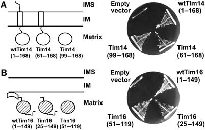
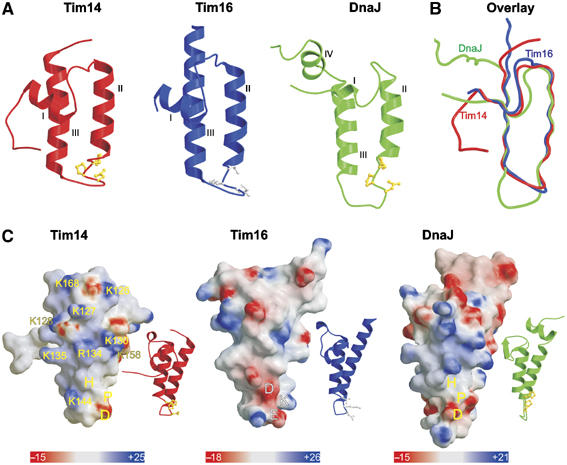
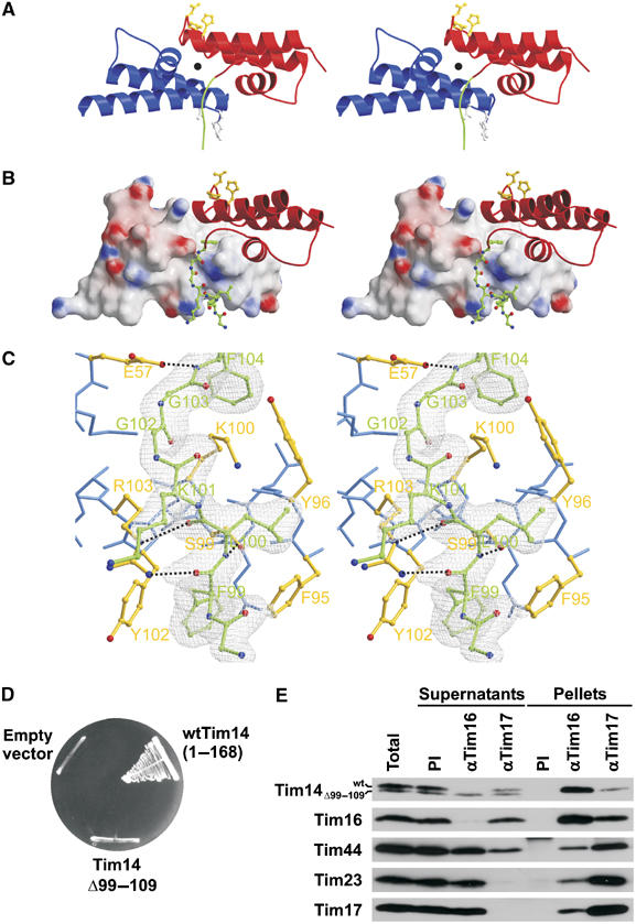
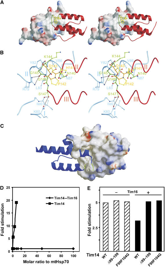
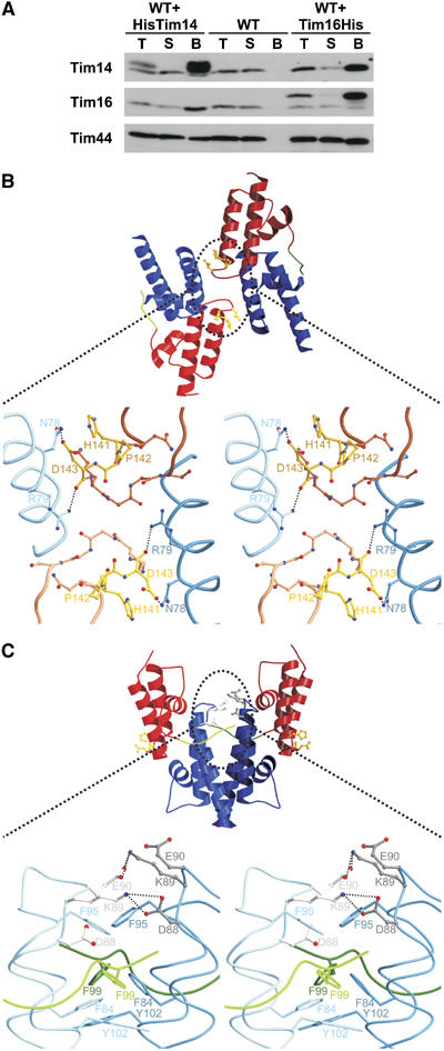
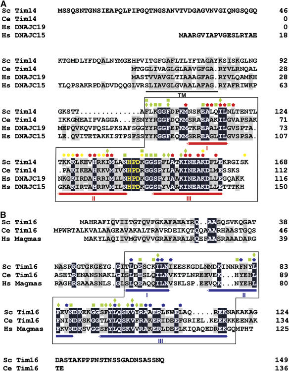
The import motor of the mitochondrial translocase of the inner membrane (TIM23) mediates the ATP-dependent translocation of preproteins into the mitochondrial matrix by cycles of binding to and release from mtHsp70. An essential step of this process is the stimulation of the ATPase activity of mtHsp70 performed by the J cochaperone Tim14. Tim14 forms a complex with the J-like protein Tim16. The crystal structure of this complex shows that the conserved domains of the two proteins have virtually identical folds but completely different surfaces enabling them to perform different functions. The Tim14-Tim16 dimer reveals a previously undescribed arrangement of J and J-like domains. Mutations that destroy the complex between Tim14 and Tim16 are lethal demonstrating that complex formation is an essential requirement for the viability of cells. We further demonstrate tight regulation of the cochaperone activity of Tim14 by Tim16. The first crystal structure of a J domain in complex with a regulatory protein provides new insights into the function of the mitochondrial TIM23 translocase and the Hsp70 chaperone system in general.


Figures

Figure 1
Figure 1
Domain analysis of Tim14 and Tim16. (A) Schematic representation of Tim14 domain structure and of truncation mutants (left panel). A haploid deletion strain of TIM14 harboring a wild-type copy of TIM14 on the URA plasmid was transformed with centromeric plasmids carrying either wild-type Tim14 or the indicated Tim14 mutants. Cells were plated on medium containing 5-fluoroorotic acid, which selects for cells that have lost the URA plasmid. Plasmids carrying wild-type Tim14 or an empty plasmid were used as positive and negative controls, respectively (right panel). (B) The same analysis was performed with Tim16. IM, inner membrane of mitochondria; IMS, intermembrane space.
Figure 2
Figure 2
Topology and charge patterns of Tim14 and Tim16. (A) Ribbon presentations of the J domain of Tim14 (left panel) and the J-like domain of Tim16 (central panel). For comparison, the J domain of DnaJ from E. coli is included (right panel). Helices I–III are indicated. HPD motifs in the J domains are presented as balls-and-sticks models and colored in yellow. Corresponding DKE motif in Tim16 is colored in gray. As compared to the J domain of DnaJ, Tim14 and Tim16 lack helix IV. (B) Superposition of the J and J-like domains of Tim14, Tim16 and DnaJ. (C) Surface representations of Tim14, Tim16 and DnaJ. Ribbon presentations of the same orientation are shown as insets. The domains are rotated clockwise by 80° relative to the representations in (A) to bring helix II to the front. Surface colors indicate positive (intense blue) and negative electrostatic potentials (intense red) with the scale bar giving the actual potentials in kT/e. Lys and Arg residues of Tim14 contributing to the positive surface charge distribution are indicated.
Figure 3
Figure 3
N-terminal arm of Tim14 embraces Tim16. (A) Ribbon model of Tim14–Tim16 heterodimer given in stereo representation. Tim14 is shown in red, Tim16 in blue. The HPD and DKE motifs are highlighted in yellow and gray, respectively, and shown as balls-and-sticks models. The pseudo-two-fold symmetry axis is indicated by a black full circle. The N-terminal arm of Tim14 is highlighted in green. (B) Stereo representation of interacting parts of Tim14 and Tim16. Tim 14 is shown as ribbon model, Tim16 as surface model. The N-terminal arm of Tim14 that embraces helix III of Tim16 is represented as balls-and-sticks model, carbon atoms are colored in green, oxygen atoms in red and nitrogen atoms in blue. (C) Stereo representation of a Tim14–Tim16 interacting section around the arm of Tim14. The electron density map (colored in gray), contoured from 1σ, is only displayed for the arm of Tim14 with 2FOFC coefficients. Temperature factor refinement indicates full occupancies of the whole arm. The residues of the arm of Tim14 are numbered in green according to sequence numbering. Residues of Tim16, which are in close contact with the arm of Tim14, are colored in yellow and represented as balls-and-sticks, whereas remaining residues of Tim16 are colored in blue. Hydrogen bonds between Tim14 and Tim16 are shown as black dotted lines. (D) A Tim14 mutant which contains an intact J domain but lacks the arm region cannot support growth of yeast cells in the absence of wild-type Tim14. The analysis was carried out as described in the legend to Figure 1A. (E) A Tim14 mutant which lacks the arm region does not interact with Tim16 in vivo. Mitochondria were isolated from wild-type yeast cells transformed with a plasmid carrying a Tim14 mutant lacking the arm region. They were solubilized with digitonin and subjected to immunoprecipitation with affinity-purified antibodies to Tim16 and Tim17 or preimmune serum (PI) as control. Supernatants and pellets were analyzed by SDS–PAGE and immunodecoration with the indicated antibodies.
Figure 4
Figure 4
The HPD loop of Tim14 is projecting into a groove formed by Tim16. (A) Stereo representation of Tim14 as ribbon model projecting into a groove of Tim16 represented as surface model. The HPD containing loop connecting helices II and III is embedded into a groove made up by residues of helices I and II of Tim16. (B) Network of hydrogen bonds between the HPD loop of Tim14 and helices I and II of Tim16 leads to a constrained conformation of the HPD loop. (C) DKE loop in Tim16 (ribbon model) is similarly inserted into a Tim14 groove (surface model). (D) The Tim14–Tim16 complex does not stimulate the ATPase activity of mtHsp70 in vitro, in contrast to Tim14 alone. The factor of stimulation was determined by measuring the ATPase activity of mtHsp70 in the presence of either various amounts of Tim14–Tim16 complex or of Tim14 and relating it to intrinsic activity of mtHsp70. (E) The arm region of Tim14 is critical for the regulation of Tim14 by Tim16. Stimulation of mtHsp70's ATPase activity by wild-type Tim14 (WT) or its two mutant forms, Tim14Δ99–109 and Tim14F99F104G, was determined in the absence (−) or presence (+) of Tim16. A typical experiment is shown.
Figure 5
Figure 5
The Tim14–Tim16 complex is organized as a tetramer. (A) His-tagged versions of Tim14 or of Tim16 were expressed in wild-type yeast cells. Mitochondria were isolated, solubilized with digitonin and the detergent extracts were incubated with NiNTA-agarose beads. Untransformed wild-type cells were used as a control. Fractions were analyzed by SDS–PAGE and immunodecoration using antibodies against Tim14, Tim16 and Tim44 as indicated. Total (T) and supernatant (S) fractions represent 20% of the material present in the bound fractions. (B) Ribbon model of type I of Tim14–Tim16 heterodimer interactions observed in the crystal. In this type of tetramer, the HPD motifs of both Tim14 are masked. The enlargement of the contact area in the stereo representation shows that the contact is made essentially by two pairs of amino-acid residues in Tim14 and Tim16. (C) Ribbon model of type II of Tim14–Tim16 heterodimer interactions in the crystal structure. The contact is made by dimerization of the Tim16 subunits of two heterodimers. The stereo representation highlights the network of hydrogen bonds. In addition, a number of hydrophobic interactions stabilize this contact.
Figure 6
Figure 6
Evolutionary conservation of Tim14 and Tim16. Sequence alignment of Tim14 (A) and Tim16 (B) with their homologues. Identical residues are shown as white letters on black background and similar residues are shaded in gray. The characteristic α helices of the J domain fold are indicated below the alignments. The HPD motif is shown in yellow. Predicted transmembrane domain (TM) of Tim14 is underlined. Red/blue pentagons—Tim14/Tim16 residues forming the hydrophobic cores of the J/J-like domains; Yellow circles—Tim14 residues which form the positively charged surface for interaction with mtHsp70; green squares/diamonds—residues involved in polar/hydrophobic interactions between Tim14 and Tim16. The parts of Tim14 and Tim16, which were crystallized, are boxed.

References

    1. Berjanskii M, Riley M, Van Doren SR (2002) Hsc70-interacting HPD loop of the J domain of polyomavirus T antigens fluctuates in ps to ns and micros to ms. J Mol Biol 321: 503–516 - PubMed
    1. Boeke JD, Trueheart J, Natsoulis G, Fink GR (1987) 5-Fluoroorotic acid as a selective agent in yeast molecular genetics. Methods Enzymol 154: 164–175 - PubMed
    1. Bradford MM (1976) A rapid and sensitive method for quantitation of microgram quantities of protein utilising the principle of protein-dye binding. Anal Biochem 72: 248–254 - PubMed
    1. Bukau B, Horwich AL (1998) The Hsp70 and Hsp60 chaperone machines. Cell 92: 351–366 - PubMed
    1. Chacinska A, Lind M, Frazier AE, Dudek J, Meisinger C, Geissler A, Sickmann A, Meyer HE, Truscott KN, Guiard B, Pfanner N, Rehling P (2005) Mitochondrial presequence translocase: switching between TOM tethering and motor recruitment involves Tim21 and Tim17. Cell 120: 817–829 - PubMed

Publication types

MeSH terms

Substances