

doi: 10.1371/journal.pcbi.0020122.
Epub 2006 Jul 28.
Intricate knots in proteins: Function and evolution
Affiliations
- PMID: 16978047
- PMCID: PMC1570178
- DOI: 10.1371/journal.pcbi.0020122
Item in Clipboard
Intricate knots in proteins: Function and evolution
PLoS Comput Biol.
.
Abstract
Our investigation of knotted structures in the Protein Data Bank reveals the most complicated knot discovered to date. We suggest that the occurrence of this knot in a human ubiquitin hydrolase might be related to the role of the enzyme in protein degradation. While knots are usually preserved among homologues, we also identify an exception in a transcarbamylase. This allows us to exemplify the function of knots in proteins and to suggest how they may have been created.
Conflict of interest statement
Figures
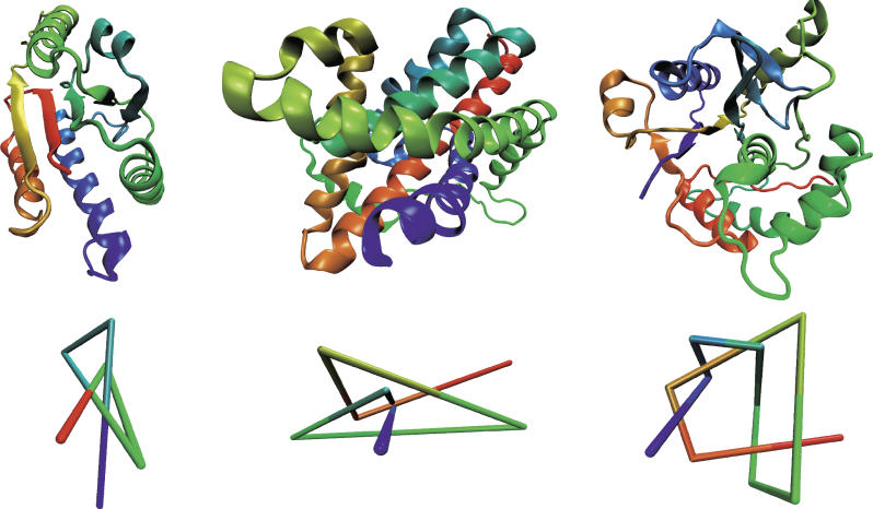
Colors change continuously from red (first residue) to blue (last residue). A reduced representation of the structure, based on the algorithm described in [1,6,36], is shown in the lower row. (Left) The trefoil knot (31) in the YBEA methyltransferase from E. coli (pdb code 1ns5; unpublished data) reveals three essential crossings in a projection onto a plane. (Middle) The figure-eight knot (41) in the Class II ketol-acid reductoisomerase from Spinacia oleracea (pdb code 1yve [26]) features four crossings. (Only the knotted section of the protein is shown.) (Right) The knot 52 in ubiquitin hydrolase UCH-L3 (pdb code 1xd3 [18]) reveals five crossings. Pictures were generated with Visual Molecular Dynamics (http://www.ks.uiuc.edu/Research/vmd ) [43].
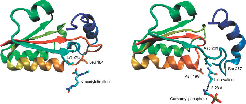
(Left) Knotted section (residues 171–278) of N-acetylornithine transcarbamylase from X. campestris with reaction product N-acetylcitrulline (pdb code 1yh1 [17]) and interacting side chains. (Right) Corresponding (unknotted) section (residues 189–286) in human ornithine transcarbamylase (pdb code 1c9y [31]) with inhibitor L-norvaline and carbamyl phosphate. Colors change continuously from red (first residue in the section) to blue (last residue in the section). The two proteins have an overall sequence identity of 29% [41]. Pictures were generated with VMD [43].
References
-
- Virnau P, Kantor Y, Kardar M. Knots in globule and coil phases of a model polyethylene. J Am Chem Soc. 2005;127:15102–15106. - PubMed
-
- Mansfield ML. Knots in Hamilton cycles. Macromolecules. 1994;27:5924–5926.
-
- Lua RC, Borovinskiy AL, Grosberg AY. Fractal and statistical properties of large compact polymers: A computational study. Polymer. 2004;45:717–731.
-
- Mansfield ML. Are there knots in proteins? Nat Struct Mol Bio. 1994;1:213–214. - PubMed
-
- Mansfield ML. Fit to be tied. Nat Struct Mol Bio. 1997;4:166–167. - PubMed
Publication types
MeSH terms
Substances
Associated data
- Actions
- Actions
- Actions
- Actions
- Actions
- Actions
- Actions
- Actions
- Actions
- Actions
- Actions
- Actions
- Actions
- Actions
LinkOut - more resources
Full Text Sources
Other Literature Sources
