

Brain magnetic resonance imaging and manganese concentrations in red blood cells of smelting workers: search for biomarkers of manganese exposure
- PMID: 16978697
- PMCID: PMC3983995
- DOI: 10.1016/j.neuro.2006.08.005
Brain magnetic resonance imaging and manganese concentrations in red blood cells of smelting workers: search for biomarkers of manganese exposure
Abstract
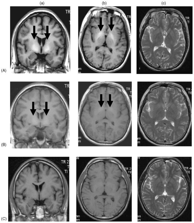
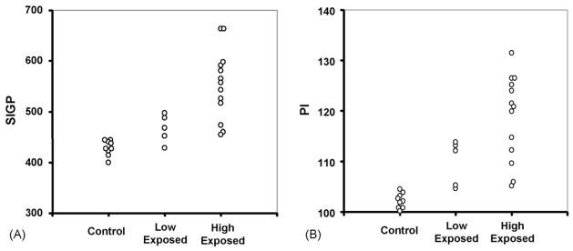
The MRI technique has been used in diagnosis of manganism in humans and non-human primates. This cross-sectional study was designed to explore whether the pallidal signal intensity in T1-weighted MRI correlated with Mn levels in the blood compartment among Mn-exposed workers and to understand to what extent the MRI signal could reflect Mn exposure. A group of 18 randomly selected male Mn-exposed workers of which 13 were smelting workers with high exposure (mean of airborne Mn in work place: 1.26 mg/m3; range: 0.31-2.93 mg/m3), and 5 power distribution control workers with low exposure (0.66 mg/m3 and 0.23-0.77 mg/m3) from a ferroalloy factory, and another group of 9 male subjects as controls from a non-smelting factory who were office or cafeteria workers (0.01 mg/m3 and 0-0.03 mg/m3) were recruited for neurological tests, MRI examination, and analysis of Mn in whole blood (MnB), plasma (MnP) or red blood cells (MnRBC). No clinical symptoms and signs of manganism were observed among these workers. MRI data showed average increases of 7.4% (p<0.05) and 16.1% (p<0.01) in pallidal index (PI) among low- and high-exposed workers, respectively, as compared to controls. Fourteen out of 18 Mn-exposed workers (78%) had intensified PI values, while this proportion was even higher (85%) among the high Mn-exposed workers. Among exposed workers, the PI values were significantly associated with MnRBC (r=0.55, p=0.02). Our data suggest that the workers exposed to airborne Mn, but without clinical symptoms, display an exposure-related, intensified MRI signal. The MRI, as well as MnRBC, may be useful in early diagnosis of Mn exposure.
Figures

References
-
- Alves G, Thiebot J, Tracqui A, Delangre T, Guedon C, Lerebours E. Neurologic disorders due to brain manganese deposition in a jaundiced patient receiving long-term parenteral nutrition. J Parenter Enteral Nutr. 1997;21:41–5. - PubMed
-
- Andrews NC. The iron transporter DMT1. Int J Biochem Cell Biol. 1999;31:991–4. - PubMed
-
- Arona A, Mata M, Bonet M. Diagnosis of chronic manganese intoxication by magnetic resonance imaging. New Engl J Med. 1997;336:964–5. - PubMed
-
- Aschner M, Vrana KE, Zheng W. Manganese uptake and distribution in the central nervous system (CNS) Neurotoxicology. 1999;20:173–80. - PubMed
-
- Butterworth RF, Spahr L, Fontaine S, Layrargues GP. Manganese toxicity, dopaminergic dysfunction and hepatic encephalopathy. Metab Brain Dis. 1995;10:259–67. - PubMed
Publication types
MeSH terms
Substances
Grants and funding
LinkOut - more resources
Full Text Sources
Medical
Research Materials
Miscellaneous
