. 2007 Feb 5;358(1):119-35.
doi: 10.1016/j.virol.2006.07.053. Epub 2006 Sep 18.

CD4 and MHC-I downregulation are conserved in primary HIV-1 Nef alleles from brain and lymphoid tissues, but Pak2 activation is highly variable

Affiliations

CD4 and MHC-I downregulation are conserved in primary HIV-1 Nef alleles from brain and lymphoid tissues, but Pak2 activation is highly variable

Kristin Agopian et al. Virology. .

Abstract

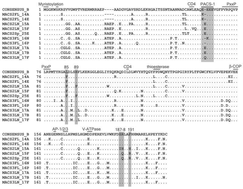
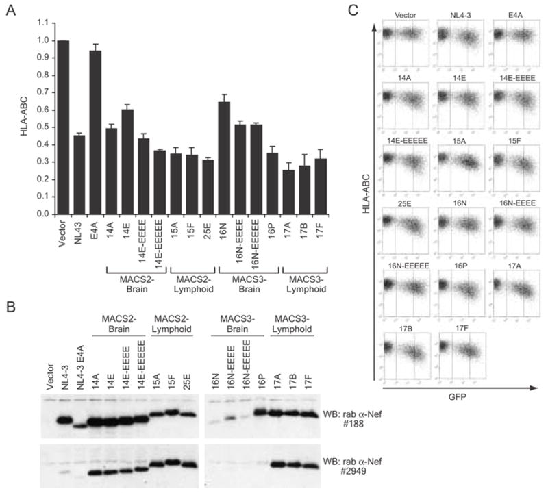
HIV-1 compartmentalization in the CNS has been demonstrated for gag, pol, and env genes. However, little is known about tissue compartmentalization of nef genes and their functional characteristics in brain. We have cloned 97 nef genes and characterized 10 Nef proteins from autopsy brain and lymphoid tissues from 2 patients with AIDS and HIV-1-associated dementia. Distinct compartmentalization of brain versus lymphoid nef genes was demonstrated within each patient. CD4 and MHC-I downregulation were conserved in all tissue-derived Nefs. However, MHC-I downregulation by brain-derived Nefs was weaker than downregulation by lymphoid-derived Nefs. The motifs KEEE- or EKEE- at the PACS-1 binding site represented brain-specific signature patterns in these 2 patients and contributed to the reduced MHC-I downregulation activity of brain-derived Nefs from these patients. Pak2 association was highly variable in Nefs from both patients. Three of 10 tissue-derived Nefs coimmunoprecipitated activated Pak2, with strong association demonstrated for only 2 Nefs. The ability of Nef to associate with activated Pak2 did not correlate with brain or lymphoid tissue origin. Nef genes from viruses isolated from brain by coculture with PBMC were not closely related to sequences amplified directly from brain tissue, suggesting that viral selection or adaptation occurred during coculture. This study of tissue-derived HIV-1 Nefs demonstrates that CD4 and MHC-I downregulation are highly conserved Nef functions, while Pak2 association is variable in late stage AIDS patients.


Figures

Figure 1
Figure 1. Phylogenetic analysis of full-length HIV-1 nef nucleotide sequences from autopsy tissue samples and viral isolates from AIDS patients MACS2 (A) and MACS3 (B)
Nef sequences amplified directly from frontal lobe, spleen, and lymph node tissues are color-coded pink, green and black, respectively. Nef sequences from brain and lymph node viral isolates obtained by PBMC co-culture are color-coded orange and blue, respectively. *MACS2-br isolate clones all contained a frameshift resulting in truncation after 60 amino acids and undetectable protein expression. To determine the phylogenetic relationship of viral isolate MACS2-br to other MACS2 sequences, the entire region of the MACS2-br 4G sequence corresponding to the intact nef ORF was used in the alignment. Numbers associated with each branch are bootstrap values, which represent the number of trees, out of 1000 replicates performed, in which the same branching order was found. Only values above 700 for the major branches are shown. Branch lengths are proportional to amount of sequence divergence. Scale bars indicate 1% sequence divergence. An ellipse indicates tissue-derived clones used in functional analyses. Isolate-derived clones analyzed previously (Agopian et al., 2006) are indicated by a rectangle.
Figure 2
Figure 2. Amino acid alignment of primary HIV-1 Nef clones with the Clade B consensus
The PACS-1 binding site, important for MHC downregulation (Piguet et al., 2000), and residues 85, 89, 187, 188 and 191, important for Pak2 association (Agopian et al., 2006; O’Neill et al., 2006) are shaded. Bars mark the locations of other proposed Nef domains, including the myristoylation signal (Geyer et al., 2001), the PxxP SH3-binding region (Saksela et al., 1995), the putative CD4 binding site (Grzesiek et al., 1996), and the putative binding sites for thioesterase (Cohen et al., 2000), and the endocytic machinery components β-COP (Piguet et al., 1999a), adaptor proteins AP-1/2/3 (Bresnahan et al., 1998; Craig et al., 1998; Greenberg et al., 1998a) and vacuolar ATPase (Lu et al., 1998). The Clade B consensus is from the LANL sequence database (August 2004).
Figure 3
Figure 3. Downregulation of cell surface CD4 is conserved in primary Nef alleles from brain and lymphoid tissues
(A) Quantitation of flow cytometric analysis of cell surface CD4 in HIJ cells co-transfected with pCDNA3-EGFP and either pCR3.1 (Vector) or pCR3.1-Nef plasmids as indicated. Relative surface CD4 was calculated from the geometric mean PE fluorescence in GFP-positive cells as described in Materials and Methods and is shown relative to GFP-positive vector-transfected cells. Averages are from 3 independent experiments. Error bars represent standard deviations. (B) Nef expression in HIJ cells assayed for CD4 downregulation is confirmed by Western blot. Lysates were split and identical membranes were blotted in parallel with either rabbit anti-Nef #188 (raised to Bru) or rabbit anti-Nef #2949. (C) Primary FACS data for flow cytometric analysis of cell surface CD4 in transfected HIJ cells. Vertical lines indicate the threshold for GFP positive cells. Data shown are representative of three independent experiments.
Figure 4
Figure 4. Downregulation of cell surface MHC-I by primary Nef proteins from brain and lymphoid tissues
(A) Quantitation of flow cytometric analysis of cell surface HLA-ABC in Jurkat T-Antigen cells co-transfected with pCDNA3-EGFP and either pCR3.1 (Vector) or pCR3.1-Nef plasmids as indicated. Relative surface HLA-ABC was calculated from the geometric mean PE fluorescence in GFP-high-expressing cells as described in Materials and methods and is shown relative to GFP-high vector-transfected cells. Averages are from 2 to 5 independent experiments. Error bars represent standard deviations. (B) Nef expression in Jurkat T-Antigen cells used for MHC-I downregulation assays confirmed by Western blot. Lysates were split and identical membranes were blotted in parallel with either rabbit anti-Nef #188 (raised to Bru) or rabbit anti-Nef #2949. (C) Primary FACS data for flow cytometric analysis of cell surface MHC-I in transfected Jurkat T-Antigen cells. Vertical lines indicate the thresholds for GFP-negative, GFP-low and GFP-high expressing cells. Data shown are representative of two to five independent experiments.
Figure 5
Figure 5. Association of primary HIV-1 Nef proteins with activated Pak2 is highly variable
(A) In vitro kinase assay (IVKA). 293T cells transiently expressing GFP and the indicated Nef proteins were immunoprecipitated with sheep-anti-Nef (raised to SF2 Nef) and assayed by IVKA (top panel) as described in Materials and methods. SF2 Nef and MACS2 5C Nef, previously demonstrated to strongly activate Pak2 (Arora et al., 2000)and (Agopian et al., 2006), are included as positive controls. An arrowhead indicates the 62 kDa band corresponding to Pak2. Whole cell lysates used for IVKA immunoprecipitations were immunoblotted with rabbit-anti-Nef antibody #2949 or sheep anti-Nef (bottom panels). (B) 293T cells transiently expressing GFP and the indicated Nef proteins were immunoprecipitated with sheep-anti-Nef and Western blotted with rabbit anti-Nef #2949 to confirm efficient Nef immunoprecipitation (top panel). Input whole cell lysates were immunoblotted with anti-Nef antibody #2949 (middle panel) or anti-GFP (bottom panel).
Figure 6
Figure 6. Phylogenetic analysis of HIV-1 Nef amino acid sequences from AIDS patients MACS2 (A) and MACS3 (B)
(A) MACS2 Nef proteins containing the F89I187H188H191 motif are colored black. Nef clones containing other motifs at these residues are colored blue. (B) MACS3 Nef proteins containing the Clade B consensus phenylalanine at residue 90 are colored black and Nef clones with a leucine at position 90 are colored blue. Bootstrap values above 700 (out of 1000) are shown. Scale bars represent 1% amino acid sequence divergence. Ellipses or rectangles indicate clones analyzed for CD4 and MHC-I downregulation and Pak2 association in this study or previously (Agopian et al., 2006).

References

    1. Agopian K, Wei BL, Garcia JV, Gabuzda D. A Hydrophobic Binding Surface on the Human Immunodeficiency Virus Type 1 Nef Core Is Critical for Association with p21-Activated Kinase 2. J Virol. 2006;80(6):3050–61. - PMC - PubMed
    1. Aiken C, Krause L, Chen YL, Trono D. Mutational analysis of HIV-1 Nef: identification of two mutants that are temperature-sensitive for CD4 downregulation. Virology. 1996;217(1):293–300. - PubMed
    1. Alexander L, Du Z, Rosenzweig M, Jung JU, Desrosiers RC. A role for natural simian immunodeficiency virus and human immunodeficiency virus type 1 nef alleles in lymphocyte activation. J Virol. 1997;71(8):6094–9. - PMC - PubMed
    1. Arold ST, Baur AS. Dynamic Nef and Nef dynamics: how structure could explain the complex activities of this small HIV protein. Trends Biochem Sci. 2001;26(6):356–63. - PubMed
    1. Arora VK, Molina RP, Foster JL, Blakemore JL, Chernoff J, Fredericksen BL, Garcia JV. Lentivirus Nef specifically activates Pak2. J Virol. 2000;74(23):11081–7. - PMC - PubMed

Publication types

MeSH terms

Substances

Associated data