. 2006 Oct 20;363(2):482-95.
doi: 10.1016/j.jmb.2006.08.063. Epub 2006 Aug 26.

Structural basis of interaction between urokinase-type plasminogen activator and its receptor

Affiliations

Structural basis of interaction between urokinase-type plasminogen activator and its receptor

Cyril Barinka et al. J Mol Biol. .

Abstract

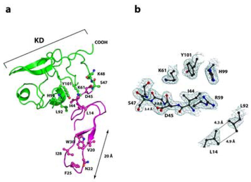
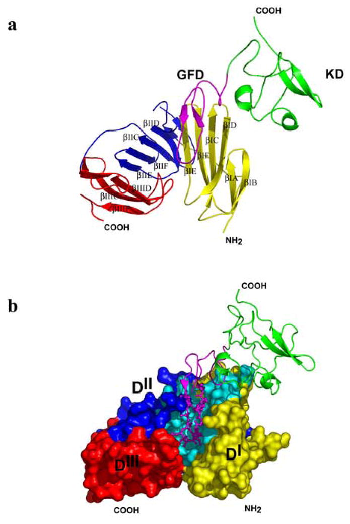
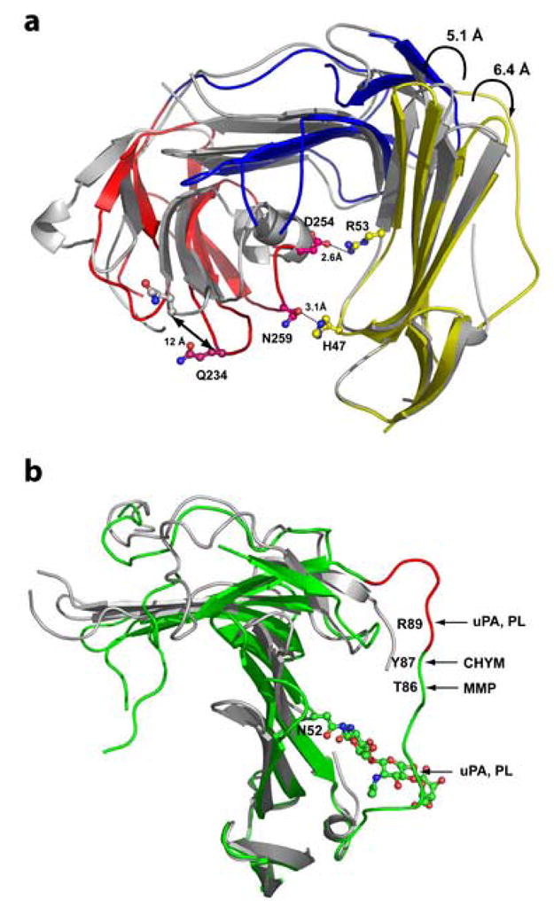
Recent studies indicate that binding of the urokinase-type plasminogen activator (uPA) to its high-affinity receptor (uPAR) orchestrates uPAR interactions with other cellular components that play a pivotal role in diverse (patho-)physiological processes, including wound healing, angiogenesis, inflammation, and cancer metastasis. However, notwithstanding the wealth of biochemical data available describing the activities of uPAR, little is known about the exact mode of uPAR/uPA interactions or the presumed conformational changes that accompany uPA/uPAR engagement. Here, we report the crystal structure of soluble urokinase plasminogen activator receptor (suPAR), which contains the three domains of the wild-type receptor but lacks the cell-surface anchoring sequence, in complex with the amino-terminal fragment of urokinase-type plasminogen activator (ATF), at the resolution of 2.8 A. We report the 1.9 A crystal structure of free ATF. Our results provide a structural basis, represented by conformational changes induced in uPAR, for several published biochemical observations describing the nature of uPAR/uPA interactions and provide insight into mechanisms that may be responsible for the cellular responses induced by uPA binding.


Figures

Figure 1
Figure 1. Overall structure of unbound ATF
Panel A: The growth-factor-like domain (GFD, amino acids 10–46) is shown in magenta and the kringle domain (KD, amino acids 47–132), in green. The Ω-loop (amino acids 19–31) is responsible for the high-affinity interactions between uPA and uPAR. Amino acids Trp30, Ile28, Phe25, Asn22 and Val20, which form a hydrophobic patch within the Ω-loop that interacts primarily with the domain DI of uPAR, are shown in ball-and-stick representation. Ball-and-stick representation is also used to highlight amino acid residues forming the inter-domain interface between GFD and KD. The atoms are colored blue (nitrogen), red (oxygen), or according to the domain-color (carbons). Panel B: The representative electron density at the interface of GFD and KD. Interactions between amino acid residues implicated in inter-domain contacts between GFD and KD constrain the ATF in an invariant shape that is virtually identical for both unbound and receptor engaged ATF. Nitrogen atoms are blue, oxygen red, carbon atoms black. The 2Fo-Fc difference electron density map is contoured at the 1.2σ level. The picture was generated using Molscript, Bobscript and rendered with PovRay.
Figure 2
Figure 2. Schematic representation of ATF binding to uPAR
Domains DI (amino acids 1–93), DII (amino acids 94–191) and DIII (amino acids 192–277) of suPAR are shown in yellow, blue, and red, respectively; KD (47–132) is in green, GFD (10–46) in magenta. Panel A: A cartoon representation of the suPAR2345/ATF complex. The individual βstrands of suPAR2345 are labeled according to refs.,. Domain DI: βIA (residues 2–8), βIB (13–17), βIC (24–33), βID (38–45), βIE (53–59), and βIF (63–70); domain DII: βIIA (94–100), βIIB (112–115), βIIC (122–129), βIID (143–149), βIIE (156–161), and βIIF (163–171); domain DIII: βIIIA (195–199), βIIIB (211–214), βIIIC (222–229), βIIID (236–243), and βIIIE (259–267). Contacts between the domains are mediated via interactions βIE and βIID (domains DI and DII), βIIE and βIIID (domains DII and DIII). Panel B: The ATF (cartoon representation) binds to the central cavity of suPAR2345 (surface representation) and the Ω-loop (Cys19-Cys31, ball-and-sticks) is primarily responsible for the high affinity binding. Residues of suPAR2345 interacting with ATF are in cyan.
Figure 3
Figure 3. Detailed view of amino acid residues implicated in ATF binding in the individual domains DI (A), and DII (B) of suPAR2345
Isolated domains DI (1–77) and DII (94–177) are shown in cartoon representation and side chains of amino acids interacting with ATF (shown in magenta, amino acids 17–41) are shown as ball-and-sticks and are colored cyan (carbon), red (oxygen), and blue (nitrogen). Panel A: The DI-ATF interaction is stabilized mostly by the hydrophobic contacts involving 16 amino acid residues from domain the DI. Panel B: Amino acid residues within domain DII interacts primarily with Lys23 and Tyr24 of ATF (shown in magenta as balls-and-sticks). Additional interactions include contacts between the fragment Pro138-Asp140, which is a part of the uPAR loop implicated in integrin signaling, and the residues Cys19-Lys23 of the Ω-loop in ATF.
Figure 4
Figure 4. Binding of ATF induces substantial conformational changes in the suPAR
The domains DI (amino acids 1–77) of the suPAR2345/ATF and suPAR/AE147 (RCSB PDB code 1YWH) complexes were aligned using corresponding Cα atoms. Panel A: Ribbon representation of the receptors (ligands were omitted for clarity) with structurally aligned domains DI. Domains DI, DII and DIII of the suPAR2345/ATF complex are in shown in yellow, blue, and red, respectively, whereas receptor from the complex suPAR/AE147 is painted in gray. The binding of ATF results in the displacement of the domain the DIII (red) by more than 12 Å and shifts the domains DI and DIII close to each other , enabling a ‘closure’ of the suPAR. Interaction between DI and DIII is mediated by two pairs of residues, His47-Asn259 and Arg53-Asp254 (shown in ball-and-stick representation). Other structural changes include repositioning of hairpins βIC-βID, βIE-βIF as well as βIIC-βIID. Panel B: Displacement of the linker region (amino acid residues Gln78-Tyr92), connecting domains DI and DII of suPAR upon ATF binding (suPAR2345/ATF structure in green, 1YWH in grey). The epitope with chemotactic attributes (amino acids Ser88-Tyr92) is show in red and positions susceptible to hydrolysis by various proteases are indicated with arrows. The trisaccharide chain, attached to Asn52 and modeled in our structure, is also shown in ball-and-stick representation. Note that “typical” mammalian oligosaccharide structures are much larger and due to their flexibility could easily “shield” the linker region from a proteolytic attack. PL, uPA, MMP, and CHYM stand for plasmin, urokinase-type plasminogen activator, matrix metallopeptidases, and chymotrypsin, respectively. The domain DIII was omitted for clarity.
Figure 5
Figure 5. Repositioning of the “integrin-interacting” loop (Trp129-Arg142) in suPAR2345 upon ATF binding
Interactions between amino acid residues Cys19-Lys23 of the ATF and Pro138-Asp140 of suPAR2345 leads to bending of the loop towards the central cavity of the receptor. Panel A: The complexes, suPAR2345/ATF (domains DI, DII and DIII colored yellow, blue and red, respectively) and suPAR/AE147 (shown in grey), were aligned based on corresponding Cα atoms of the domain DI only. GFD is shown in combination of ball-and-sticks and semi-transparent surface. The βIIC-βIID hairpin is in cartoon representation and its residues interacting with GFD as ball-and-sticks. Panel B: Detailed view of residues engaged in the interactions between the strands βIIC and βIID of suPAR2345 and the Ω-loop of ATF. The Ω-loop is shown in surface representation and the interacting residues contributed by the domain DII are painted as balls-and-sticks. Note the major movement of the βIIC-βIID hairpin caused by interactions with the Ω-loop. In both structures amino acids 132 through 136 of suPAR are missing.
Figure 6
Figure 6. Binding of ATF in the central cavity of suPAR differs from the inhibitory peptide AE147
The complexes suPAR2345/ATF and suPAR/AE147 were superimposed using the equivalent Cα atoms of the receptors. The AE147 peptide (green) and ATF (fragment Cys13-Cys31, magenta) are in cartoon representation. In the ball-and-stick representation are shown the residues marked in bold in the FXXYLW (AE147) or KYFXXIHW (ATF) motifs. The residues of receptor forming the binding pocket are displayed in surface representation. The labels of residues in AE147 are printed in blue and slanted, those of the ATF residues are shown in red, and the suPAR residues are printed in black. Due to presence of resembling sequence motifs (FXXXLW), similar binding modes were proposed for the ATF and AE147. From this figure, it is clear that despite of a spatial overlap between the two ligands, except of Phe25 (ATF) and Phe5 (AE147), locations of the individual side chains differ markedly.

Similar articles

Cited by

References

    1. Mondino A, Blasi F. uPA and uPAR in fibrinolysis, immunity and pathology. Trends Immunol. 2004;25:450–455. - PubMed
    1. Romer J, Nielsen BS, Ploug M. The urokinase receptor as a potential target in cancer therapy. Curr Pharm Des. 2004;10:2359–2376. - PubMed
    1. Rabbani SA, Mazar AP. The role of the plasminogen activation system in angiogenesis and metastasis. Surg Oncol Clin N Am. 2001;10:393–415. - PubMed
    1. Czekay RP, Kuemmel TA, Orlando RA, Farquhar MG. Direct binding of occupied urokinase receptor (uPAR) to LDL receptor-related protein is required for endocytosis of uPAR and regulation of cell surface urokinase activity. Mol Biol Cell. 2001;12:1467–1479. - PMC - PubMed
    1. Degryse B, Resnati M, Czekay RP, Loskutoff DJ, Blasi F. Domain 2 of the urokinase receptor contains an integrin-interacting epitope with intrinsic signaling activity: generation of a new integrin inhibitor. J Biol Chem. 2005;280:24792–24803. - PubMed

Publication types

MeSH terms

Substances