. 2006 Nov 17;281(46):35156-66.
doi: 10.1074/jbc.M606409200. Epub 2006 Sep 15.

Loss of CNGB1 protein leads to olfactory dysfunction and subciliary cyclic nucleotide-gated channel trapping

Affiliations

Loss of CNGB1 protein leads to olfactory dysfunction and subciliary cyclic nucleotide-gated channel trapping

Stylianos Michalakis et al. J Biol Chem. .

Abstract

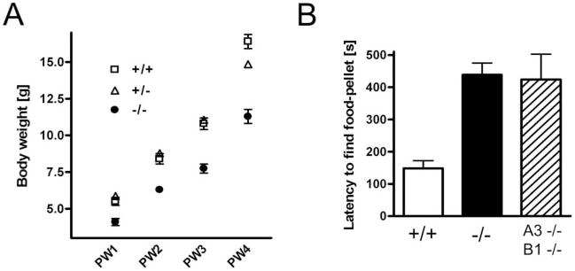
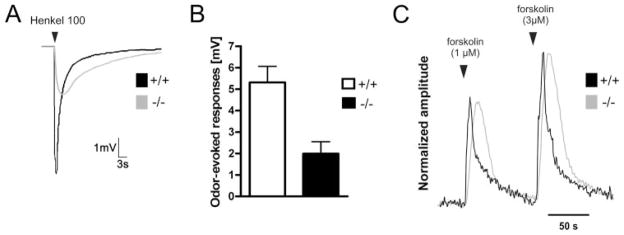
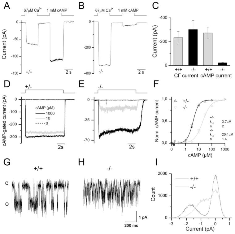
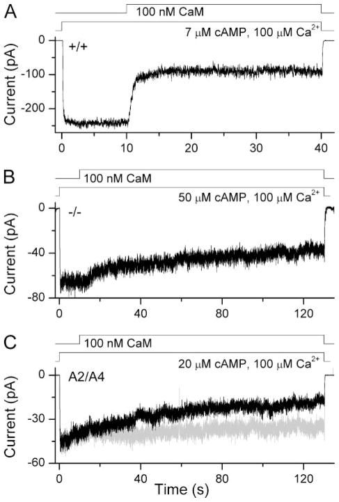
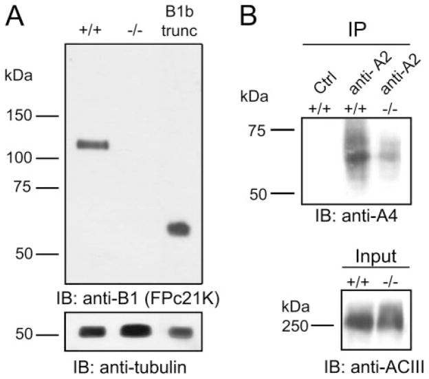
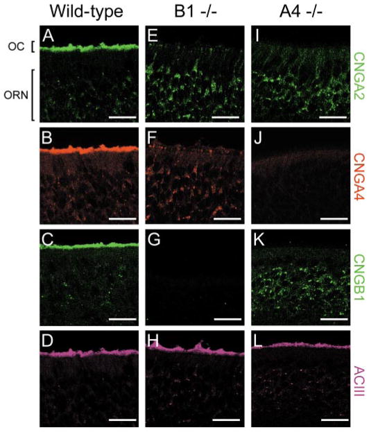
Olfactory receptor neurons (ORNs) employ a cyclic nucleotide-gated (CNG) channel to generate a receptor current in response to an odorant-induced rise in cAMP. This channel contains three types of subunits, the principal CNGA2 subunit and two modulatory subunits (CNGA4 and CNGB1b). Here, we have analyzed the functional relevance of CNGB1 for olfaction by gene targeting in mice. Electro-olfactogram responses of CNGB1-deficient (CNGB1-/-) mice displayed a reduced maximal amplitude and decelerated onset and recovery kinetics compared with wild-type mice. In a behavioral test, CNGB1-/- mice exhibited a profoundly decreased olfactory performance. Electrophysiological recordings revealed that ORNs of CNGB1-/- mice weakly expressed a CNG current with decreased cAMP sensitivity, very rapid flicker-gating behavior and no fast modulation by Ca2+-calmodulin. Co-immunoprecipitation confirmed the presence of a CNGA2/CNGA4 channel in the olfactory epithelium of CNGB1-/- mice. This CNGA2/CNGA4 channel was targeted to the plasma membrane of olfactory knobs, but failed to be trafficked into olfactory cilia. Interestingly, we observed a similar trafficking defect in mice deficient for the CNGA4 subunit. In conclusion, these results demonstrate that CNGB1 has a dual function in vivo. First, it endows the olfactory CNG channel with a variety of biophysical properties tailored to the specific requirements of olfactory transduction. Second, together with the CNGA4 subunit, CNGB1 is needed for ciliary targeting of the olfactory CNG channel.


Figures

FIGURE 1
FIGURE 1. Delayed postnatal body weight increase and reduced olfactory performance in CNGB1−/− mice
A, body weight curves of littermate CNGB1+/+ (open squares; n = 14 –28), CNGB1+/− (open triangles; n = 14 – 82), and CNGB1−/− (filled circles; n = 12–58) mice from the first postnatal week (PW) to PW4. The data for female and male mice were pooled. B, CNGB1−/− and CNGA3−/−/CNGB1−/− double knock-out mice reveal a strongly increased mean latency to localize a hidden food pellet in an olfactory task.
FIGURE 2
FIGURE 2. Morphology of the olfactory epithelium of wild-type and CNGB1−/− mice
A, toluidine blue-stained vertical semithin sections through the olfactory epithelium (turbinate IV) of CNGB1+/+ and CNGB1−/− mice (litter-matched, 10-week-old). B, vertical sections through the olfactory epithelium of CNGB1+/+ and CNGB1−/− mice immunostained with an antibody specific for the olfactory marker protein. Scale bars are 10 μm (A) and 20 μm (B).
FIGURE 3
FIGURE 3. The olfactory bulb of CNGB1−/− mice is smaller than that of wild-type mice but reveals functional activity
A, gross brain morphology of litter-matched 4-week-old CNGB1+/+ and CNGB1−/− mice. The white lines indicate the length of the wild-type olfactory bulb. B, coronal sections through the olfactory bulb of CNGB1+/+and CNGB1−/− mice stained with anti-tyrosine hydroxylase antibody. C, odor-induced Fos expression in periglomerular neurons in CNGB1+/− and CNGB1−/− mice after incubation of the mice with fresh air (left panel) or air-saturated with a 100-compound odor mixture (Henkel 100) (right panel). The arrowheads point to knock-out glomeruli displaying Fos expression. D, quantification of total and Fos-positive (Fos+) glomeruli in slices from identical regions of the olfactory bulb of CNGB1+/+ (white bars; n = 3) and CNGB1−/− (black bars; n = 3) mice. Whereas the total number of glomeruli and the number of Fos+ glomeruli per slice is smaller in CNGB1−/− mice the fraction of Fos+ glomeruli (%Fos+) is identical between both genotypes. ns, not significant. *, p < 0.05. Scale bars are 2 mm (A), 200 μm (B), and 50 μm (C).
FIGURE 4
FIGURE 4. Significantly altered odor responses in CNGB1−/− mice
A, representative EOG traces of CNGB1+/+ (black) and CNGB1−/− (gray) mice. Responses were obtained after a 1-s pulse of Henkel 100. B, mean peak amplitudes of Henkel 100-evoked responses were significantly smaller in CNGB1−/− (black bar; n = 15) than in CNGB1+/+mice (white bar; n = 10) (p < 0.005). C, representative traces of ratiofluorometric recordings from CNGB1+/+ (black) and CNGB1−/− (gray) ORN knobs revealed delayed Ca2+ -signals in knock-out mice. Traces were normalized to the maximum response allowing comparison of response kinetics.
FIGURE 5
FIGURE 5. The olfactory CNG channel from CNGB1−/− ORNs reveals a reduced peak-current amplitude, a lower cAMP sensitivity, and displays an extremely rapid flicker-gating behavior
Patches excised from dendritic knobs of CNGB1+/+ (A) and CNGB1−/− (B) ORNs were exposed to 67 μM Ca2+ or 1 mM cAMP. Because the Cl current exhibits “rundown” over time, traces were recorded within 15 s after patch excision. C, mean Cl and CNG currents in patches from CNGB1+/+ and CNGB1−/− mice. Cl current: CNGB1+/+ 232 ± 53 pA; CNGB1−/−, 301 ± 76 pA (n.s.; n = 9). CNG current: CNGB1+/+, 273 ± 49 pA; CNGB1b−/−, 23 ± 3 pA (p < 0.005; n = 9). D and E, currents induced by 10 and 1000 μM cAMP, respectively, from an excised, inside-out membrane patch from the dendritic knob of a CNGB1+/− (D) and a CNGB1−/− (E) ORN. F, normalized cAMP dose response relationships from CNGB1+/− and CNGB1−/− mice. Currents were determined 10 s after exposure to cAMP. Solid lines represent Hill curves fitted to the data. G–I, single channel activity elicited in patches excised from CNGB1+/+ (G) and CNGB1−/− (H) ORNs by applying 3 and 10 μM cAMP, respectively. Open (o) and closed (c) levels are indicated in the figure. I, all-point amplitude histogram of 18-s exposures to cAMP of channels from CNGB1+/+ (black trace) and CNGB1−/− (gray trace) ORNs. All experiments were performed in symmetrical NaCl solution at a holding potential of −40 mV (A–F) or −60 mV (G–I).
FIGURE 6
FIGURE 6. Slow inactivation of the CNG channel by Ca2+-CaM in CNGB1−/− ORNs
Patches excised from CNGB1+/+ (A), CNGB1−/− (B) ORNs or HEK293 cells transfected with CNGA2 and CNGA4 (C) were exposed to levels of cAMP, chosen to yield an open probability of 0.75 (7, 50, and 20 μM cAMP, respectively). Application of Ca2+ -CaM quickly reduced the CNG current only in patches from CNGB1+/+ ORNs, while channels from CNGB1−/− or heterologously expressed CNGA2/CNGA4 channels showed a similarly slow inactivation. In C the gray trace represents the current evoked by cAMP in the absence of Ca2+ -CaM. All experiments were performed in symmetrical Na-methanesulfonate solution, and the pipette solution contained 1 mM niflumic acid to suppress the Ca2+ -activated Cl current.
FIGURE 7
FIGURE 7. Detection of a CNGA2/CNGA4 channel in CNGB1−/− olfactory epithelium
A, top panel: immunoblot of membrane fractions from olfactory tissue of CNGB1+/+ or CNGB1−/− mice probed with an antibody directed against the CNGB1b N terminus (FPc21K). Membrane fractions of HEK293 cells expressing a truncated CNGB1b protein (B1b trunc) representing the hypothetical gene product encoded by the knock-out-CNGB1 locus are loaded in the right lane. Bottom panel, same blot probed with anti-tubulin antibody. B, top panel, CNGA2 co-immunoprecipitates with CNGA4 in CNGB1+/+and CNGB1−/− mice. Equal amounts of membranes from olfactory tissue (210 μg) were precipitated with rabbit anti-CNGA2 (anti-A2) or a control antibody (anti-PAK, Ctrl), respectively, blotted, and probed with guinea pig anti-CNGA4 (anti-A4). Bottom panel, loading control representing 15% of the membranes from olfactory tissue used for the co-immunoprecipitation shown in the top panel of B probed with rabbit anti-adenylyl cyclase III (ACIII).
FIGURE 8
FIGURE 8. Analysis of CNG channel expression in wild-type and mutant olfactory epithelium
Confocal images of sections from 4-week-old wild-type (WT) (A–D), CNGB1−/− (E–H), and CNGA4−/− (I–L) mice labeled with antibodies specific for CNGA2, CNGA4, CNGB1, and the adenylyl cyclase (ACIII). For detection of CNGB1 an antibody specific for the C terminus was used. Scale bar represents 20 μm. The position of the olfactory cilia (OC) and the ORN cell bodies are marked with brackets in A. Images in panels (A, B, and D), (E, F, and H), and (J, K, and L) were obtained from triple staining using the indicated antibodies.
FIGURE 9
FIGURE 9. Inhibition of the proteasome increases the total CNGA2 protein level in ORNs of CNGB1−/− mice but does not promote targeting of the protein to the cilia
Immunostaining showing the expression of CNGA2 and ACIII in the olfactory epithelium of litter-matched 4-week-old CNGB1−/− mice 20 h after intraperitoneal injection with buffer (A–C), with a single dose (10 mg/g) of the proteasome inhibitor MG-132 (D–F) or with two doses of MG-132 applied at consecutive days (G–I). Scale bar is 10 μm. The position of the olfactory cilia (OC) and the olfactory knobs (OK) are marked with brackets. All images are projections of confocal z-stacks.

Similar articles

Cited by

References

    1. Nakamura T, Tsuru K, Miyamoto S. Neurosci Lett. 1994;171:197–200. - PubMed
    1. Frings S, Seifert R, Godde M, Kaupp UB. Neuron. 1995;15:169–179. - PubMed
    1. Kurahashi T, Yau KW. Nature. 1993;363:71–74. - PubMed
    1. Kleene SJ, Gesteland RC. J Neurosci. 1991;11:3624–3629. - PMC - PubMed
    1. Lowe G, Gold GH. Nature. 1993;366:283–286. - PubMed

Publication types

Substances