. 2006 Nov;174(3):1635-59.
doi: 10.1534/genetics.106.061283. Epub 2006 Sep 15.

A gain-of-function screen identifying genes required for vein formation in the Drosophila melanogaster wing

Affiliations

A gain-of-function screen identifying genes required for vein formation in the Drosophila melanogaster wing

Cristina Molnar et al. Genetics. 2006 Nov.

Abstract

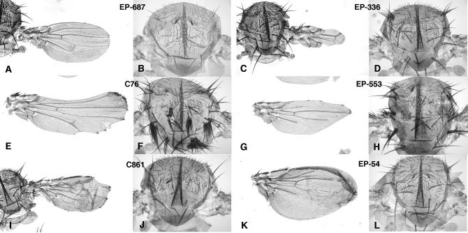
The formation of the Drosophila wing involves developmental processes such as cell proliferation, pattern formation, and cell differentiation that are common to all multicellular organisms. The genes controlling these cellular behaviors are conserved throughout the animal kingdom, and the genetic analysis of wing development has been instrumental in their identification and functional characterization. The wing is a postembryonic structure, and most loss-of-function mutations are lethal in homozygous flies before metamorphosis. In this manner, loss-of-function genetic screens aiming to identify genes affecting wing formation have not been systematically utilized. As an alternative, a number of genetic searches have utilized the phenotypic consequences of gene gain-of-expression, as a method more efficient to search for genes required during imaginal development. Here we present the results of a gain-of-function screen designed to identify genes involved in the formation of the wing veins. We generated 13,000 P-GS insertions of a P element containing UAS sequences (P-GS) and combined them with a Gal4 driver expressed mainly in the developing pupal veins. We selected 500 P-GSs that, in combination with the Gal4 driver, result in modifications of the veins, changes in the morphology of the wing, or defects in the differentiation of the trichomes. The P-element insertion sites were mapped to the genomic sequence, identifying 373 gene candidates to participate in wing morphogenesis and vein formation.


Figures

F<sc>igure</sc> 1.—
Figure 1.—
Gal4 and P-UAS lines used in the screen, schematic of the genetic crosses, and modifications to the vein pattern resulting from modifications in the Notch, EGFR, and Dpp signaling pathways during pupal development. (A) Expression of green fluorescent protein (GFP) in Gal4-shv3Kpn/UAS-GFP pupal wings 36 hr APF. (B) Higher magnification of the vein L3 showing expression of GFP in the cell membranes of vein cells and expression of Blistered (Bs) in the nucleus of intervein cells. (C) Tangential section of B showing the complementary domains of Bs and GFP expression in the intervein and vein, respectively. (D) Representation of the Gal4-shvkpn vector. (E) Representation of the P-GS vector showing the UAS sequences and Hsp70b promoter near the inverted terminal repeats (IR) at both P-element ends. The mini-white gene (white) and the position of the restriction sites used to map the P-GS insertions (HhaI and MspI) are indicated in E. (F) Generation of new P-GS insertions using Δ2-3 transposase to mobilize a P-GS element inserted on a CyO chromosome. The flies with a novel P-GS insertion were crossed to Gal4-shv3kpn flies to induce the expression of the genes adjacent to the vector, and flies with a wing mutant phenotype were selected to establish balanced lines. (G) Ectopic expression in the pupal veins of a dominant-negative form of Notch (Gal4-shv3Kpn/UAS-Necd; −N) results in strong thickening of the veins. (H) Ectopic expression of a dominant-negative form of EGFR (Gal4-shv3Kpn/UAS-EGFRDN; −EGFR) causes loss of veins. (I) Expression of the Dpp-antagonist Dad (Gal4- shv3Kpn/UAS-dad; −Dpp) causes loss of veins. (J) Expression of the intracellular fragment of Notch (Gal4- shv3Kpn/UAS-Nintra; +N) eliminates the veins. (K) Expression of an activated form of the EGFR downstream component Ras (Gal4- shv3Kpn/UAS-RasV12; +EGFR) causes the formation of thicker veins. (L) Expression of the ligand Dpp (Gal4- shv3Kpn/UAS-dpp; +Dpp) causes the differentiation of most wing tissue as vein.
F<sc>igure</sc> 2.—
Figure 2.—
Numerical parameters of the screen. (A) Genomic sites identified grouped by the number of P-GS insertions located in a similar position (±1 kb) in the molecular map. Most genomic sites have been identified by only one insertion (149). (B) Distance of P-GS insertions to the closest adjacent gene. Most of the insertions are situated within a gene or at a distance of <1 kb (183). (C) Frequency of phenotypic classes in the combinations between P-GS insertions and Gal4-shv3kpn: 30% of insertion sites result in thicker veins (V+), 16% produce loss of veins (V−), 22% cause alterations in trichome differentiation (CD), 20% cause defects in dorsal–ventral apposition (B) and 12% result in misfolded wings (F).
F<sc>igure</sc> 3.—
Figure 3.—
Representative phenotypes obtained in the combinations of P-GS lines with Gal4-shv3kpn. (A–C) Loss of vein differentiation: strong phenotype (A, Gal4-shv3kpn/EP-160), weak phenotype (B, Gal4-shv3kpn/C502) and loss of the L4 vein (C, Gal4-shv3kpn/C495). (D–F) Vein thickening: strong phenotype (D, Gal4-shv3kpn/EP-600), weak phenotype (E, Gal4-shv3kpn/C474) and thickened L3 vein (F, Gal4-shv3kpn/EP-738). (G) “Blistered” or dorsal–ventral apposition phenotype (Gal4-shv3kpn/C547). (H) Folded wing (Gal4-shv3kpn/C56). (I) Vein thickening and loss of trichome differentiation phenotype (Gal4-shv3kpn/EP-167). (J–M) Higher magnification of the L3 vein showing cell differentiation defects such as loss of trichomes (J, Gal4-shv3kpn/C464), differentiation of several trichomes per cell (K, Gal4-shv3kpn/C125), loss of trichomes and reduced pigmentation (L, Gal4-shv/C386), and formation of smaller than normal trichomes (M, Gal4-shv3kpn/C432).
F<sc>igure</sc> 4.—
Figure 4.—
Effects of P-GS insertion on adjacent genes. (A) Schematic of the genomic region where the P-GS insertions EP-33 and C861 (triangles) are localized, showing the annotated transcription units as horizontal thick solid arrows. (B–E) In situ hybridization with Stat92E (B), CG5180 (C), and att (D) antisense probes and with att sense probe (E) in EP-33/Gal4-sal wing imaginal discs. Ectopic expression in the spalt domain is detected only for Stat92E (B) and CG5180 (C) and no expression is detected for the transcript oriented “antisense” with respect to the insertion site (att in D). (F–K) In situ hybridization with Stat92E (F), att (G), CG5180 (H), and CG5191 (K) antisense probes and with sense probes for the transcripts CG15922 (I) and CG10877 (J) in C861/Gal4-sal wing imaginal discs. Ectopic expression is only detected for the transcripts att (G) and CG5180 (H) and no Gal4-driven expression is detected for the transcript with antisense orientation (CG15922 in I and CG10877 in J). (L) Schematic of the genomic region where the P-GS insertion EP-866 (triangle) is localized. (M–Q) In situ hybridization with CG9056 (M), CG9066 (O), CG15916 (P), and shi (Q) antisense probes and with CG9066 sense probe (N) in EP-866/Gal4-sal wing imaginal discs. Ectopic expression is detected for the three transcripts oriented “sense” with respect the insertion site (CG9056, CG15916, and shi), whereas no expression is detected for the transcript with antisense orientation (CG9066).
F<sc>igure</sc> 5.—
Figure 5.—
Comparison between the phenotype of P-GS insertion and UAS constructs. (A–H) Representative examples of P-GS/Gal4 (A–D) and UAS/Gal4 (E–H) combinations. (A and E) Partial loss of L3 vein caused by ectopic expression of hh in pupal veins using the P-GS line C279 (A) and UAS-hh (E). (B and F) Similar loss of vein differentiation caused by ectopic expression of dad in pupal veins using the P-GS line EP-459 (B) and UAS-dad (F). (C and G) Differentiation of extra-vein tissue by ectopic expression of dpp in pupal veins using the P-GS line C517 (C) and UAS-dpp (G). (D and H) Loss of veins and reduced wing size observed when hairy is expressed ectopically in the wing imaginal disc using the P-GS line C107 (D) and UAS-h (H).
F<sc>igure</sc> 6.—
Figure 6.—
Representative phenotypes observed in combinations between P-GS insertions and the Gal4 lines 638 and 253. The phenotypes in the wing (A, C, E, G, I, and K) in combinations with Gal4-638 and thorax (B, D, F, H, J, and L) in combinations with Gal4-253 of the P-GS insertions EP-687 (A and B), EP-336 (C and D), C76 (E and F), EP-553 (G and H), C861 (I and J), and EP-54 (K and L) are shown. The effects in the wing consist of loss of veins (A, G, and K), ectopic vein tissue (I), thickened veins (E), and loss of wing margin and associated wing tissue (A, C, E, G, and I). The phenotypes in the thorax are loss of macrochaetae (B, J, and L) and ectopic macrochaetae in clusters (D, F, and H).
F<sc>igure</sc> 7.—
Figure 7.—
Frequency of phenotypic classes obtained in combinations between the P-GS lines and the Gal4 lines Gal4-638 (A, C, E, G, and I) and Gal4-253 (B, D, F, H, and J). The P-GS lines have been grouped after the phenotypes resulting in combinations with Gal4-shv3Kpn: thicker veins (A and B), loss of veins (C and D), cell differentiation (E and F), blistered (G and H), and folded wing (I and J). (A–D) Most P-GS lines affecting the veins in combination with Gal4-shv3Kpn also affect the veins when the ectopic expression is induced in the wing disc with Gal4-638 (A and C). A high percentage of P-GS insertions causing thicker veins in combination with Gal4-shv3Kpn also produce defects in the wing margin in combination with Gal4-638, whereas many P-GS insertions causing loss of veins in combination with Gal4-shv3Kpn eliminate the macrochaetae in combination with Gal4-253 (C and D, respectively). In contrast, many P-GS lines causing wing blistering and folded wing phenotypes in combination with Gal4-shv3Kpn (G and I, respectively) result in wild-type phenotypes in combinations with both Gal4-638 and Gal4-253 (H and J). Symbols: V+, ectopic or thicker veins; V−, loss of veins; S, reduced wing size; S-P, reduced wing size and altered vein pattern; N, notched wings; nW, absence of wing blade tissue; B, blistered wing; F, folded wing; L, larval or pupal lethal; E, epithelial integrity alterations; wt, normal wings and normal chaetae pattern; +Mq, extra-macrochaetae; −Mq, loss of macrochaetae.
F<sc>igure</sc> 8.—
Figure 8.—
Molecular classification of the genes identified grouped in the phenotypic classes obtained in combination with Gal4-shv: thicker veins (A), loss of veins (B), cell differentiation (C), blistered (D), and folded wing (E). The more represented molecular classes correspond to genes encoding proteins involved in cell signaling (CS) and transcription factors (TF), particularly for candidate genes causing loss of veins (B). Symbols: CS, signaling molecules; TF, transcription factors; M, metabolism; CA, cell adhesion; CY, cytosqueleton components; RB, RNA binding proteins; PP, protein proteases; CG, computer-annotated genes without identified structural domains; CGh, computer-annotated genes with vertebrate homologs; CGd, computer-annotated genes with a structural motive; snRNA, small nuclear RNA; microRNA, microRNA; tRNA, tRNA.
F<sc>igure</sc> 9.—
Figure 9.—
Schematic of the core components belonging to the EGFR (A), Notch (B), and Dpp (C) signaling pathways. The relationships between different pathway members are indicated with arrows to denote activation or with bars to indicate repression. The genes included within shaded squares were identified in the screening as P-GS insertions close to their 5′ transcription start.

References

    1. Abdelilah-Seyfried, S., Y. M. Chan, C. Zeng, N. J. Justice, S. Younger-Shepherd et al., 2000. A gain-of-function screen for genes that affect the development of the Drosophila adult external sensory organ. Genetics 155: 733–752. - PMC - PubMed
    1. Adams, M. D., S. E. Celniker, R. A. Holt, C. A. Evans, J. D. Gocayne et al., 2000. The genome sequence of Drosophila melanogaster. Science 287: 2185–2195. - PubMed
    1. Adler, P. N., 2002. Planar signaling and morphogenesis in Drosophila. Dev. Cell 2: 525–535. - PubMed
    1. Babcock, M. C., R. S. Stowers, J. Leither, C. S. Goodman and L. J. Pallanck, 2003. A genetic screen for synaptic transmission mutants mapping to the right arm of chromosome 3 in Drosophila. Genetics 165: 171–183. - PMC - PubMed
    1. Bier, E., 2000. Drawing lines in the Drosophila wing: initiation of wing vein development. Curr. Opin. Genet. Dev. 10: 393–398. - PubMed

Publication types