. 2006 Nov;174(3):1189-204.
doi: 10.1534/genetics.106.057604. Epub 2006 Sep 15.

The large isoform of Drosophila melanogaster heterochromatin protein 2 plays a critical role in gene silencing and chromosome structure

Affiliations

The large isoform of Drosophila melanogaster heterochromatin protein 2 plays a critical role in gene silencing and chromosome structure

Christopher D Shaffer et al. Genetics. 2006 Nov.

Abstract

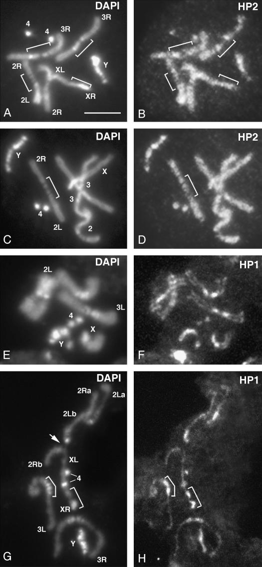
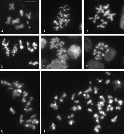
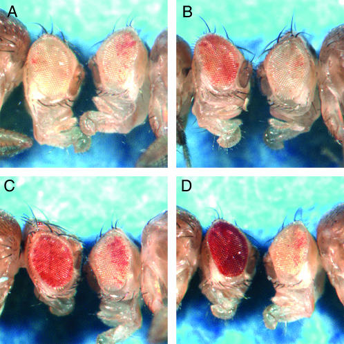
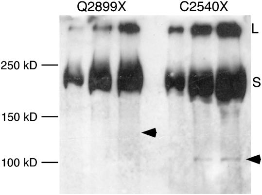
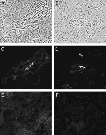
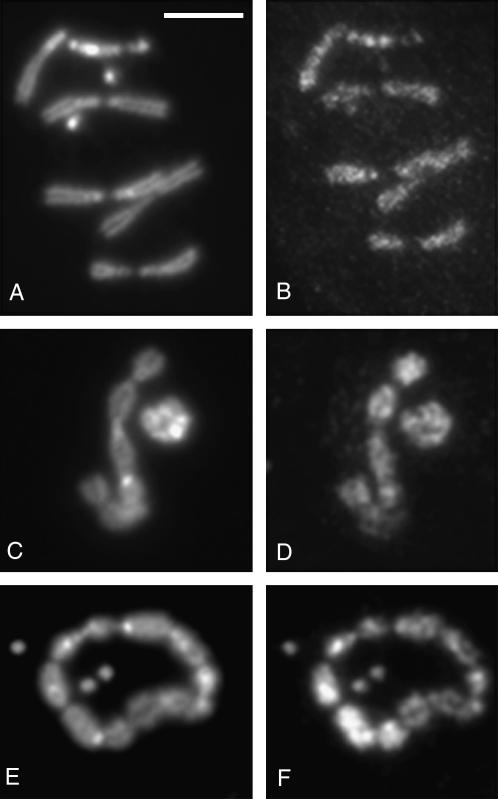
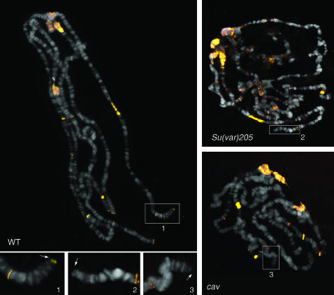
Drosophila melanogaster heterochromatin protein 2 (HP2) interacts with heterochromatin protein 1 (HP1). In polytene chromosomes, HP2 and HP1 colocalize at the chromocenter, telomeres, and the small fourth chromosome. We show here that HP2 is present in the arms as well as the centromeric regions of mitotic chromosomes. We also demonstrate that Su(var)2-HP2 exhibits a dosage-dependent modification of variegation of a yellow reporter transgene, indicating a structural role in heterochromatin formation. We have isolated and characterized 14 new mutations in the Su(var)2-HP2 gene. Using wm4h, many (but not all) mutant alleles show dominant Su(var) activity. Su(var)2-HP2 mutant larvae show a wide variety of mitotic abnormalities, but not the telomere fusion seen in larvae deficient for HP1. The Su(var)2-HP2 gene codes for two isoforms: HP2-L (approximately 365 kDa) and HP2-S (approximately 175 kDa), lacking exons 5 and 6. In general, mutations that affect only the larger isoform result in more pronounced defects than do mutations common to both isoforms. This suggests that an imbalance between large and small isoforms is particularly deleterious. These results indicate a role for HP2 in the structural organization of chromosomes and in heterochromatin-induced gene silencing and show that the larger isoform plays a critical role in these processes.


Figures

F<sc>igure</sc> 1.—
Figure 1.—
Immunolocalization of HP2 and HP1 along prometaphase chromosomes of wild-type larval brains. Two examples are shown. (A, C, E, and G) DAPI-stained chromosomes. (B, D, F, and H) Chromosomes immunostained for either HP2 (B and D) using a rabbit antiserum (Shaffer et al. 2002) or HP1 (F and H) using the C1A9 monoclonal (James and Elgin 1986). Characteristic heterochromatic regions, designated by brackets, were identified by their DAPI-banding pattern using the cytological map of Drosophila heterochromatin (Gatti and Pimpinelli 1992). In addition, the Y chromosome and the fourth (“dot”) chromosome are largely heterochromatic. In F and H, one of the second chromosomes is bent, so that its 2La and 2Ra arms lie parallel; the other second chromosome displays a prominent secondary constriction (arrow) that corresponds to the histone gene cluster. See text for further explanation. Bar, 5 μm.
F<sc>igure</sc> 2.—
Figure 2.—
HP2 dosage affects variegated yellow expression. Dissected abdomens from four different D. melanogaster lines carrying different doses of Su(var)2-HP2 were scored for yellow expression from the variegating transgene B079. (A and B) The yellow expression level of flies carrying the two parental chromosomes used to generate the Df(2R)B11 and Dp(2R)214B chromosomes. These two lines each carry two wild-type copies of the gene for HP2. (C) The yellow expression level of a fly carrying one copy of the gene for HP2, generated using Df(2R)B11; note the increased number of pigmented patches. (D) The yellow expression level of a fly carrying three copies of the gene for HP2, generated using Dp(2R)214B; note the decreased number of pigmented patches.
F<sc>igure</sc> 3.—
Figure 3.—
Summary of the Su(var)2-HP2 alleles by type and location. Each exon is indicated by a large rectangle; the shaded exons are the HP2-L-specific exons, and the solid exons are common to both HP2-L and HP2-S (HP2-L, top line; HP2-S, bottom line). The small numbers at the ends of some of the exons of the larger isoform indicate the number of the amino acid found at the exon boundary. The open rectangles labeled “A” and “T” indicate the location of the two AT hooks found within HP2-L. The open oval labeled “H” indicates the position of the HP1-binding domain (Stephens et al. 2005). The location of each mutation described is indicated with a solid arrow. Arrows above the larger isoform and pointing downward indicate the position of the three missense alleles. The arrows and half-arrows below the isoforms and pointing upward indicate the locations of the truncation alleles. The number at the end of the arrow indicates the level of suppression of variegation, from near complete (3) to no (0) loss of silencing (see Figure 5). The letter above or below the Su(var) number indicates the extent of mitotic abnormalities: S, very frequent abnormalities; M, medium level of abnormalities; N, no abnormalities (see Table 2). The amino acid change for allele 288 is T588I, the amino acid change for 230 is N3220I, and allele 692 is a single-base insertion of an A, which produces a frameshift at amino acid 2370. The amino acid change for all other alleles is indicated by the allele name.
F<sc>igure</sc> 4.—
Figure 4.—
Chromosome abnormalities in Su(var)2 -HP2 mutants. (A) DAPI-stained wild-type male metaphase chromosomes. (B–H) Examples of chromosome abnormalities observed in Su(var)2-HP2 mutants. (B–D) Metaphase chromosomes from Su(var)2-HP2Q2259X/Df(2R)B11 (B and C) and Su(var)2-HP2Q895X/Df(2R)B11 (D) mutant brains displaying poorly condensed chromosomes. (E) Metaphase chromosomes from Su(var)2-HP2Q2259X/Df(2R)B11 mutants displaying PSCS. (F) Su(var)2-HP2Q1187X/Df(2R)B11 mutant displaying strongly condensed chromosomes and PSCS. (G and H) Polyploid mutant metaphases from Su(var)2-HP2Q770X/Df(2R)B11 (G) and Su(var)2-HP2Q2097X/Df(2R)B11 (H) mutant brains with poorly condensed chromosomes displaying PSCS. Bar, 5 μm.
F<sc>igure</sc> 5.—
Figure 5.—
Eye phenotypes of Su(var)2-HP2 alleles show a range in suppression of PEV. Males carrying a wm4h X chromosome are shown. In each picture, the left fly carries a Su(var)2-HP2 mutation, whereas the right fly is a sibling carrying a wild-type copy of Su(var)2-HP2. Alleles were separated into four classes as shown: (A) class 0, no suppression, example Su(var)2-HP2Q2540X; (B) class 1, weak suppression, example Su(var)2-HP2Q1187X; (C) class 2, medium suppression, example Su(var)2-HP2Q895X; and (D) class 3, strong suppression, example Su(var)2-HP2Q770X. Quantitative assessment of these example lines showed an effect that ranged from no apparent suppression (OD480 indistinguishable from wm4h homozygotes at 7–9% of wild-type pigment) (classes 0 and 1) to ∼17 and 31% of wild-type pigment (classes 2 and 3) with ∼2% standard error for triplicate samples.
F<sc>igure</sc> 6.—
Figure 6.—
Stable truncated products from the HP2 mutations are generally not observed in a Western blot. Proteins extracted from isolated nuclei from females heterozygous for the indicated Su(var)2-HP2 allele (over a wild-type allele) were separated by SDS gel electrophoresis, blotted, and stained for HP2 using the HP2 chicken antiserum (which recognizes the first 15 amino acids of HP2, common to both HP2-L and HP2-S). Three lanes were loaded with increasing amounts of the protein extract in each case. The arrowhead indicates the predicted position of the truncated HP2-S protein. The numbers on the left indicate the position of several molecular weight markers. The letters on the right indicate the position of the full-length HP2-L (L) and HP2-S (S) protein products.
F<sc>igure</sc> 7.—
Figure 7.—
Heterochromatin association of HP1 is not dependent on HP2. Polytene chromosomes from HP2-depleted larvae [carrying HP2 mutant alleles over Df(2R)B11] are shown. (A, C, and E) Polytene chromosomes from a Su(var)2-HP2P2763L/Df(2R)B11 larva; (B, D, and F) Polytene chromosomes from a Su(var)2-HP2Q2259X/Df(2R)B11 larva. (A and B) Phase-contrast photograph of the chromosomes. (C and D) The same chromosomes stained with anti-HP1 antibodies. (E and F) The same chromosomes stained with anti-HP2 rabbit antiserum.
F<sc>igure</sc> 8.—
Figure 8.—
HP2 normally localizes on metaphase chromosomes from Su(var)205 mutant larvae. Chromosome preparations were immunostained with the anti-HP2 rabbit antiserum that recognizes both isoforms of HP2. Metaphase chromosomes from Oregon-R (A and B) and Su(var)20505/Su(var)20504 (C–F) larval neuroblasts. (A, C, and E) DAPI staining. (B, D, and F) HP2 localization. Bar, 5 μm.
F<sc>igure</sc> 9.—
Figure 9.—
HP2 is lost from telomeres in Su(var)205 and cav mutant polytene chromosomes. Chromosome preparations were immunostained with the anti-HP2 rabbit antiserum that recognizes both isoforms of HP2. Note that HP2 (yellow signals) accumulates at the chromocenter, at several euchromatic bands, and at all telomeres of wild-type chromosomes. In polytene chromosomes from Su(var)205 and cav mutants (right top and right bottom), HP2 is normally concentrated at the chromocenter and at the euchromatic bands but fails to localize at the telomeres. The inserts outline the tips of the 2L polytene chromosome arms and the arrows point to the telomeres.
F<sc>igure</sc> 10.—
Figure 10.—
HP2-L, but not HP2-S, requires HP1 for stable binding to the chromocenter of polytene chromosomes. Polytene chromosomes from HP1-depleted larvae [carrying heteroallelic combinations of Su(var)205 alleles] are shown. (A–D) Polytene chromosomes stained with C1A9 mouse anti-HP1 monoclonal antibody. (E and F) Chromosomes stained with anti-HP2 guinea pig antiserum directed against HP2-L;. (G and H) Chromosomes stained with anti-HP2 rabbit antiserum against HP2-L and HP2-S. (A and E) Wild-type chromosomes. (B, C, F, and G) Chromosomes from a Su(var)2055/Su(var)2052 larva. (D and H) Chromosomes from a Su(var)20505/Su(var)20504 larva.

Similar articles

Cited by

References

    1. Bannister, A. J., P. Zegerman, J. F. Partridge, E. A. Miska, J. O. Thomas et al., 2001. Selective recognition of methylated lysine 9 on histone H3 by the HP1 chromo domain. Nature 410: 120–124. - PubMed
    1. Basu, J., H. Bousbaa, E. Logarinho, Z. Li, B. C. Williams et al., 1999. Mutations in the essential spindle checkpoint gene bub1 cause chromosome missegregation and fail to block apoptosis in Drosophila. J. Cell Biol. 146: 13–28. - PMC - PubMed
    1. Blom, N., S. Gammeltoft and S. Brunak, 1999. Sequence and structure-based prediction of eukaryotic protein phosphorylation sites. J. Mol. Biol. 294: 1351–1362. - PubMed
    1. Cenci, G., G. Siriaco, G. D. Raffa, R. Kellum and M. Gatti, 2003. The Drosophila HOAP protein is required for telomere capping. Nat. Cell Biol. 5: 82–84. - PubMed
    1. Cleard, F., M. Delattre and P. Spierer, 1997. SU(VAR)3–7, a Drosophila heterochromatin-associated protein and companion of HP1 in the genomic silencing of position-effect variegation. EMBO J. 16: 5280–5288. - PMC - PubMed

Publication types

MeSH terms