. 2006 Sep 26;103(39):14379-84.
doi: 10.1073/pnas.0606714103. Epub 2006 Sep 15.

Transducer of regulated CREB-binding proteins (TORCs) induce PGC-1alpha transcription and mitochondrial biogenesis in muscle cells

Affiliations

Transducer of regulated CREB-binding proteins (TORCs) induce PGC-1alpha transcription and mitochondrial biogenesis in muscle cells

Zhidan Wu et al. Proc Natl Acad Sci U S A. .

Abstract

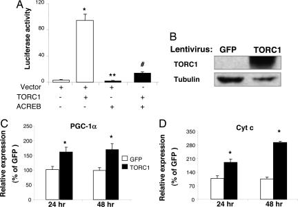
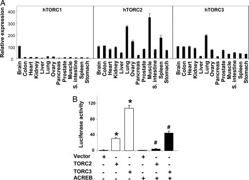
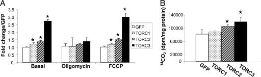
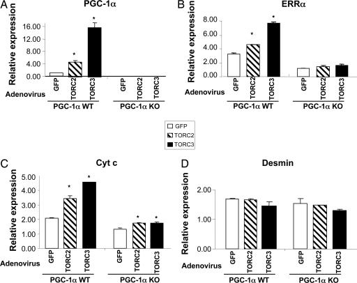
PGC-1alpha (peroxisome proliferator-activated receptor gamma coactivator 1alpha) is a master regulator of mitochondrial biogenesis and plays an important role in several other aspects of energy metabolism. To identify upstream regulators of PGC-1alpha gene transcription, 10,000 human full-length cDNAs were screened for induction of the PGC-1alpha promoter. A number of activators of PGC-1alpha transcription were found; the most potent activator was the transducer of regulated CREB (cAMP response element-binding protein) binding protein (TORC) 1, a coactivator of CREB. The other two members of the TORC family, TORC2 and TORC3, also strongly activated PGC-1alpha transcription. TORCs dramatically induced PGC-1alpha gene transcription through CREB. Forced expression of TORCs in primary muscle cells induced the endogenous mRNA of PGC-1alpha and its downstream target genes in the mitochondrial respiratory chain and TCA cycle. Importantly, these changes in gene expression resulted in increased mitochondrial oxidative capacity measured by cellular respiration and fatty acid oxidation. Finally, we demonstrated that the action of TORCs in promoting mitochondrial gene expression and function requires PGC-1alpha. Previous studies had indicated that TORCs function as a calcium- and cAMP-sensitive coincidence detector and mediate individual and synergistic effects of these two pathways. Our results, together with previous findings, strongly suggest that TORCs play a key role in linking these external signals to the transcriptional program of adaptive mitochondrial biogenesis by activating PGC-1alpha gene transcription.


Conflict of interest statement

The authors declare no conflict of interest.

Figures

Fig. 1.
Fig. 1.
TORC1 induces PGC-1α and Cyt c expression in HeLa cells. (A) HeLa cells were transfected with pGL3-basic-PGC1α-Luc (40 ng) and phRL-SV40 (3 ng, as control for transfection efficiency) along with the indicated cDNA constructs (TORC1, 60 ng; ACREB, 60 ng). The cells were lysed, and the luciferase activity was determined 48 h after transfection. The luciferase activity represents the ratio of firefly luminescence over renilla luminescence, an index of PGC-1α gene transcription; n = 6. ∗, P < 0.05 lane 2 vs. lane 1; ∗∗, P < 0.05 lane 3 vs. lane 1; #, P < 0.05 lane 4 vs. lane 2. (BD) HeLa cells were transduced with a lentivirus expressing either GFP or TORC1. Total protein was extracted from cells 48 h after transduction and subjected to Western blot analysis to determine the expression of TORC1 protein (B). Total RNA was extracted from the cells at 24 and 48 h after transduction and subjected to Q-PCR analysis to determine the expression level of endogenous PGC-1α (C) and Cyt c mRNA (D). The average relative expression (mean ± SEM) is shown. ∗, P < 0.05 TORC1 vs. GFP; n = 3.
Fig. 2.
Fig. 2.
TORC2 and TORC3 strongly induce PGC-1α transcription. (A) Human tissue distribution of the TORC family. The mRNA expression levels of TORC1, 2, and 3 were measured by Q-PCR in a panel of human tissue cDNAs. The mRNA expression level of each TORC in brain is set at 100%. The mRNA expression levels in other tissues is relative to that of brain. (B) TORC2 and TORC3 induce PGC-1α promoter activity. HeLa cells were transfected with pGL3-basic-2kb-PGC1α-Luc (40 ng) and phRL-SV40 (3 ng) along with various cDNA constructs, as indicated (TORC2 or TORC3, 10 ng; ACREB, 110 ng). The cells were lysed, and the luciferase activity was determined at 48 h after transfection. The luciferase activity represents the ratio of firefly luminescence over renilla luminescence (mean ± SEM). ∗, P < 0.05, lanes 2 and 3 vs. lane 1; #, P < 0.05, lane 5 vs. lane 2 and lane 6 vs. lane 3; n = 6
Fig. 3.
Fig. 3.
Adenoviral-mediated expression of TORCs increases mitochondrial gene expression. (A and B) Primary muscle cells were transduced with TORC1, TORC2, TORC3, or GFP adenovirus. Total protein or RNA was extracted from the cells at 48 h after transduction, and 50 μg of total protein extract from each sample was subjected to Western blot analysis to determine the expression levels of TORC1 (A), and ectopically expressed TORC2 and TORC3 protein (B). The primary antibodies used were anti-TORC1 in A and anti-V5 in B. The secondary antibody used was HRP-conjugated goat anti-rabbit IgG. Total RNA samples were subjected to Q-PCR analysis. (CG) The relative expression levels of the endogenous PGC-1α (C) and ERRα (D), Cyt c (E), Cox II (F), and IDH3α (G) are shown. Mean ± SEM; n = 3; ∗, P < 0.05, TORC1, TORC2, or TORC3 vs. GFP.
Fig. 4.
Fig. 4.
Adenoviral-mediated overexpression of TORCs increase mitochondrial oxidative capacity in muscle cells. Primary muscle cells were transduced with TORC1, TORC2, TORC3, or GFP adenovirus for 48 h. (A) Cellular respiration was measured without treatment (basal) or with treatment, as indicated. The average fold change over GFP (mean ± SEM) is shown. (B) Cells were labeled with [14C]palmitate for 2 h, and the complete fatty acid oxidation rate indicated by the amount of 14CO2 produced was determined. All experiments were performed in triplicate. ∗, P < 0.05 for TORC vs. GFP; n = 3.
Fig. 5.
Fig. 5.
The effect of TORC2 and TORC3 on mitochondrial gene expression is primarily mediated through PGC-1α. WT and PGC-1α-null primary muscle cells were transduced with TORC2, TORC3, or GFP adenovirus for 48 h. Total RNA was extracted, and the gene expression levels (mean ± SEM) were measured by Q-PCR analysis. ∗, P < 0.05, TORC2 or TORC3 vs. GFP; n = 3.

References

    1. Kelly DP, Scarpulla RC. Genes Dev. 2004;18:357–368. - PubMed
    1. Puigserver P, Wu Z, Park CW, Graves R, Wright M, Spiegelman BM. Cell. 1998;92:829–839. - PubMed
    1. Wu Z, Puigserver P, Andersson U, Zhang C, Adelmant G, Mootha V, Troy A, Cinti S, Lowell B, Scarpulla RC, Spiegelman BM. Cell. 1999;98:115–124. - PubMed
    1. Lin J, Handschin C, Spiegelman BM. Cell Metab. 2005;1:361–370. - PubMed
    1. Mootha VK, Lindgren CM, Eriksson K-F, Subramanian A, Sihag S, Lehar J, Puigserver P, Carlsson E, Ridderstrale M, Laurila E, et al. Nat Genet. 2003;34:267–273. - PubMed

MeSH terms

LinkOut - more resources