. 2006 Sep 26;103(39):14483-8.
doi: 10.1073/pnas.0605245103. Epub 2006 Sep 15.

Granulocyte colony-stimulating factor preferentially stimulates proliferation of monosomy 7 cells bearing the isoform IV receptor

Affiliations

Granulocyte colony-stimulating factor preferentially stimulates proliferation of monosomy 7 cells bearing the isoform IV receptor

Elaine M Sloand et al. Proc Natl Acad Sci U S A. .

Abstract

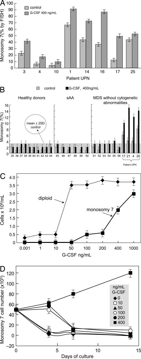
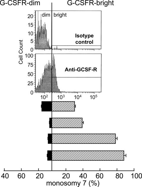
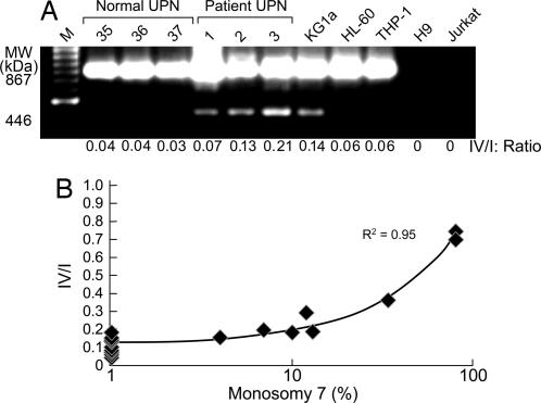
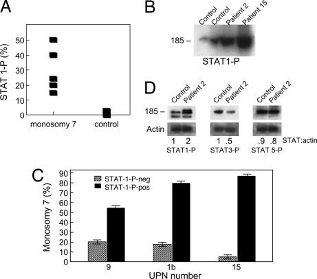
Granulocyte colony-stimulating factor (GCSF) administration has been linked to the development of monosomy 7 in severe congenital neutropenia and aplastic anemia. We assessed the effect of pharmacologic doses of GCSF on monosomy 7 cells to determine whether this chromosomal abnormality developed de novo or arose as a result of favored expansion of a preexisting clone. Fluorescence in situ hybridization (FISH) of chromosome 7 was used to identify small populations of aneuploid cells. When bone marrow mononuclear cells from patients with monosomy 7 were cultured with 400 ng/ml GCSF, all samples showed significant increases in the proportion of monosomy 7 cells. In contrast, bone marrow from karyotypically normal aplastic anemia, myelodysplastic syndrome, or healthy individuals did not show an increase in monosomy 7 cells in culture. In bone marrow CD34 cells of patients with myelodysplastic syndrome and monosomy 7, GCSF receptor (GCSFR) protein was increased. Although no mutation was found in genomic GCSFR DNA, CD34 cells showed increased expression of the GCSFR class IV mRNA isoform, which is defective in signaling cellular differentiation. GCSFR signal transduction via the Jak/Stat system was abnormal in monosomy 7 CD34 cells, with increased phosphorylated signal transducer and activation of transcription protein, STAT1-P, and increased STAT5-P relative to STAT3-P. Our results suggest that pharmacologic doses of GCSF increase the proportion of preexisting monosomy 7 cells. The abnormal response of monosomy 7 cells to GCSF would be explained by the expansion of undifferentiated monosomy 7 clones expressing the class IV GCSFR, which is defective in signaling cell maturation.


Conflict of interest statement

The authors declare no conflict of interest.

Figures

Fig. 1.
Fig. 1.
GCSF increases the proportion of monosomy 7 cells in patients with a preexisting monosomy 7 clone. BMMNC were placed in culture with and without pharmacologic doses (400 ng/ml) of GCSF for 7 days, as described in Materials and Methods. (A) All 24 patients who demonstrated monosomy 7 by conventional cytogenetics showed increased proportions and numbers of cells with monosomy 7. Results are shown for eight patients. (B) None of the 10 normal controls or 6 AA patients without clinical monosomy 7 showed a significant increase in the proportion of cells with monosomy 7 in tissue culture as a result of exposure to high GCSF doses, whereas MDS patients with monosomy 7 detectable by FISH all showed expansion of this clone after GCSF exposure. (C) A dose–response curve for BMMNC performed on three patients with monosomy 7 showing decreased sensitivity to low doses of GCSF when cultured short-term (4 days) compared with diploid cells. (D) When the cells described in C were subjected to longer culture times (14 days), monosomy 7 cells declined at lower GCSF concentrations.
Fig. 2.
Fig. 2.
GCSFR protein is increased on monosomy 7 CD34 cells. When CD34 cells from monosomy 7 patients were sorted into GCSFR-negative and -positive fractions by flow cytometry and FISH was performed, the monosomy 7 cells were predominantly in the GCSFR-positive population.
Fig. 3.
Fig. 3.
Bone marrow CD34 cells from patients with monosomy 7 express abnormal amounts of the shortened isoform IV of GCSFR. RNA from CD34 cells from monosomy 7 patients was subjected to DNase treatment (Invitrogen, Carlsbad, CA) followed by oligo(dT)-primed RT-PCR using the SuperScript First Strand synthesis system for RT-PCR (Invitrogen). GCSFR isoform amplification was performed as described in ref. . (A) Samples from three healthy volunteers, three patients, and cell lines KGi1, HL60, and THP-1, showing increased expression of shortened isoform IV. (B) When real-time PCR was performed on BMMNC, as described in Materials and Methods, an increased ratio of GCSFR-IV/I was observed that was proportional to the number of monosomy 7 cells in the bone marrow sample.
Fig. 4.
Fig. 4.
STAT P expression is abnormal in monosomy 7. Unstimulated BMMNC were stained with CD34-PE and anti-STAT1-P mAbs. (A) When CD34 cells from monosomy 7 cells not subjected to GCSF stimulation were stained with STAT1-P-FITC mAb, there was increased STAT1-P expression in patients compared with controls. (B) Constitutive expression of STAT1-P in monosomy 7 patients is shown on immunoblot. (C) When CD34 cells were sorted by flow cytometry and FISH was performed, there was a preponderance of monosomy 7 cells in the STAT1-P+ group, indicating constitutive expression of this protein in monosomy 7 cells. (D) When BMMNC from two healthy donors, four MDS patients without monosomy 7, and three patients with monosomy 7 were stimulated with GCSF (400 ng/ml) for 30 min, immunoblotting showed increased expression of STAT1-P and decreased STAT3-P (example shown).

References

    1. Young NS. Blood. 1992;79:1385–1392. - PubMed
    1. Rosenfeld R, Follman D, Nunez O, Young N. J Am Med Assoc. 2003;289:1130–1135. - PubMed
    1. Maciejewski J, Risitano A, Sloand EM, Nunez O, Young NS. Blood. 2002;99:3129–3135. - PubMed
    1. Hashino S, Imamura M, Tanaka J, Kobajashi S, Musashi M, Kasai M, Asaka M. Ann Hematol. 1996;72:337–339. - PubMed
    1. Kojima S, Ohara A, Tsuchida M, Kudoh T, Hanada R, Okimoto Y, Kaneko T, Takano T, Ikuta K, Tsukimoto I. Blood. 2002;100:786–790. - PubMed

MeSH terms