

A recA null mutation may be generated in Streptomyces coelicolor
- PMID: 16980478
- PMCID: PMC1595505
- DOI: 10.1128/JB.00951-06
A recA null mutation may be generated in Streptomyces coelicolor
Abstract
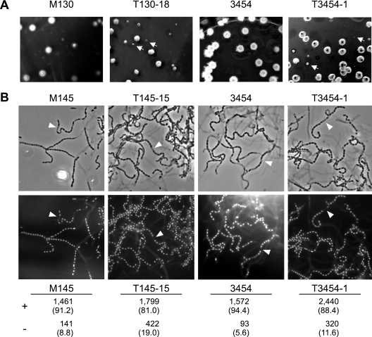
The recombinase RecA plays a crucial role in homologous recombination and the SOS response in bacteria. Although recA mutants usually are defective in homologous recombination and grow poorly, they nevertheless can be isolated in almost all bacteria. Previously, considerable difficulties were experienced by several laboratories in generating recA null mutations in Streptomyces, and the only recA null mutants isolated (from Streptomyces lividans) appeared to be accompanied by a suppressing mutation. Using gene replacement mediated by Escherichia coli-Streptomyces conjugation, we generated recA null mutations in a series of Streptomyces coelicolor A3(2) strains. These recA mutants were very sensitive to mitomycin C but only moderately sensitive to UV irradiation, and the UV survival curves showed wide shoulders, reflecting the presence of a recA-independent repair pathway. The mutants segregated minute colonies with low viability during growth and produced more anucleate spores than the wild type. Some crosses between pairs of recA null mutants generated no detectable recombinants, showing for the first time that conjugal recombination in S. coelicolor is recA mediated, but other mutants retained the ability to undergo recombination. The nature of this novel recombination activity is unknown.
Figures


Similar articles
-
Mutational analysis of the Streptomyces lividans recA gene suggests that only mutants with residual activity remain viable.Mol Gen Genet. 1997 Jul;255(4):420-8. doi: 10.1007/s004380050514. Mol Gen Genet. 1997. PMID: 9267438
-
Isolation of protease-proficient, recombinase-deficient recA mutants of Escherichia coli K-12.J Bacteriol. 1985 Aug;163(2):688-95. doi: 10.1128/jb.163.2.688-695.1985. J Bacteriol. 1985. PMID: 3160687 Free PMC article.
-
Evidence that an additional mutation is required to tolerate insertional inactivation of the Streptomyces lividans recA gene.J Bacteriol. 2001 Jul;183(14):4374-81. doi: 10.1128/JB.183.14.4374-4381.2001. J Bacteriol. 2001. PMID: 11418579 Free PMC article.
-
Isolation and characterization of spontaneous srl-recA deletion mutants in Escherichia coli K-12.Mol Gen Genet. 1984;196(2):356-9. doi: 10.1007/BF00328071. Mol Gen Genet. 1984. PMID: 6387399
-
recA mutants of E. coli K12: a personal turning point.Bioessays. 1996 Sep;18(9):767-72. doi: 10.1002/bies.950180912. Bioessays. 1996. PMID: 8831293 Review.
Cited by
-
A novel docking domain interface model predicting recombination between homoeologous modular biosynthetic gene clusters.J Ind Microbiol Biotechnol. 2011 Sep;38(9):1295-304. doi: 10.1007/s10295-010-0909-0. Epub 2010 Nov 24. J Ind Microbiol Biotechnol. 2011. PMID: 21107638
-
One-step genome engineering in bee gut bacterial symbionts.mBio. 2024 Sep 11;15(9):e0139224. doi: 10.1128/mbio.01392-24. Epub 2024 Aug 6. mBio. 2024. PMID: 39105596 Free PMC article.
-
The evolution of no-cost resistance at sub-MIC concentrations of streptomycin in Streptomyces coelicolor.ISME J. 2017 May;11(5):1168-1178. doi: 10.1038/ismej.2016.194. Epub 2017 Jan 17. ISME J. 2017. PMID: 28094796 Free PMC article.
-
Genome-Wide Identification of the LexA-Mediated DNA Damage Response in Streptomyces venezuelae.J Bacteriol. 2022 Aug 16;204(8):e0010822. doi: 10.1128/jb.00108-22. Epub 2022 Jul 13. J Bacteriol. 2022. PMID: 35862789 Free PMC article.
-
DNA polymerase I is not required for replication of linear chromosomes in streptomyces.J Bacteriol. 2008 Jan;190(2):755-8. doi: 10.1128/JB.01335-07. Epub 2007 Nov 9. J Bacteriol. 2008. PMID: 17993519 Free PMC article.
References
-
- Ahel, I., A. Mikoc, and V. Gamulin. 2005. recA gene expression in a streptomycete is mediated by the unusual C-terminus of RecA protein. FEMS Microbiol. Lett. 248:119-124. - PubMed
-
- Aigle, B., A. C. Holl, J. F. Angulo, P. Leblond, and B. Decaris. 1997. Characterization of two Streptomyces ambofaciens recA mutants: identification of the RecA protein by immunoblotting. FEMS Microbiol. Lett. 149:181-187. - PubMed
-
- Bentley, S. D., K. F. Chater, A.-M. Cerdeño-Tárraga, G. L. Challis, N. R. Thomson, K. D. James, D. E. Harris, M. A. Quail, H. Kieser, D. Harper, A. Bateman, S. Brown, G. Chandra, C. W. Chen, M. Collins, A. Cronin, A. Fraser, A. Goble, J. Hidalgo, T. Hornsby, S. Howarth, C. H. Huang, T. Kieser, L. Larke, L. Murphy, K. Oliver, S. O'Neil, E. Rabbinowitsch, M. A. Rajandream, K. Rutherford, S. Rutter, K. Seeger, D. Saunders, S. Sharp, R. Squares, S. Squares, K. Taylor, T. Warren, A. Wietzorrek, J. Woodward, B. G. Barrell, J. Parkhill, and D. A. Hopwood. 2002. Complete genome sequence of the model actinomycete Streptomyces coelicolor A3(2). Nature 417:141-147. - PubMed
-
- Capaldo, F. N., and S. D. Barbour. 1975. DNA content, synthesis and integrity in dividing and non-dividing cells of rec− strains of Escherichia coli K12. J. Mol. Biol. 91:53-66. - PubMed
Publication types
MeSH terms
Substances
LinkOut - more resources
Full Text Sources
