Comparative Study
. 2006 Sep-Oct;13(5):638-43.
doi: 10.1101/lm.251906. Epub 2006 Sep 15.

Distinct roles for medial temporal lobe structures in memory for objects and their locations

Affiliations
Comparative Study

Distinct roles for medial temporal lobe structures in memory for objects and their locations

Elizabeth A Buffalo et al. Learn Mem. 2006 Sep-Oct.

Abstract

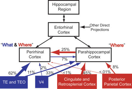
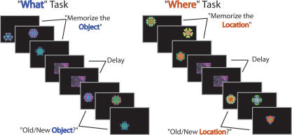
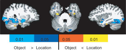
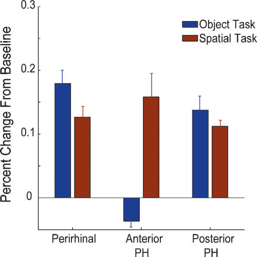
The ability to learn and retain novel information depends on a system of structures in the medial temporal lobe (MTL) including the hippocampus and the surrounding entorhinal, perirhinal, and parahippocampal cortices. Damage to these structures produces profound memory deficits; however, the unique contribution to memory of each of these structures remains unclear. Here we have used functional magnetic resonance imaging (fMRI) to determine whether the perirhinal and parahippocampal cortices show differential memory-related activity. Based on the distinct patterns of cortical input to these two areas, we reasoned that these structures might show differential activity for spatial and object recognition memory. In each of 11 subjects, we found that the perirhinal cortex was active during both spatial and object memory encoding, while the anterior parahippocampal cortex was active only during spatial encoding. These data support the idea that MTL structures make distinct contributions to recognition memory performance.


Figures

Figure 1.
Figure 1.
Cortical input to the medial temporal lobe. A schematic diagram showing the percentage of cortical input from unimodal and polymodal areas to the perirhinal and parahippocampal cortices in the medial temporal lobe (black boxes). The percentages of cortical input shown on this schematic are from Suzuki and Amaral (1994a). These data suggest that the parahippocampal cortex might be more important for spatial memory (red lines and boxes), while the perirhinal cortex might be more important for object memory (blue lines and boxes). Additionally, these data raise the possibility that the perirhinal cortex is involved in both spatial and object memory because of the large amount of spatial information it receives via the parahippocampal cortex. The thickness of the lines approximately represents the relative percentages of cortical input.
Figure 2.
Figure 2.
Task design. While being scanned, subjects were shown a series of six colored stimuli and were instructed to memorize the object (Object task) or memorize the location (Spatial task). During a delay, subjects viewed a scrambled image presented six times at the center of the screen. During the recognition phase, subjects were shown three old objects and three new objects (Object task) or objects in three old locations and three new locations (Spatial task). For each stimulus, subjects made an Old/New judgment by pressing buttons on a button box.
Figure 3.
Figure 3.
What versus where task activity. In an individual subject, the two tasks activated different areas in the visual processing pathway. (A) Areas that showed greater activation during the Object task (cool colors) or during the Location task (warm colors). (B) The respective averaged time series for these activations.
Figure 4.
Figure 4.
Task-specific encoding activity. Results from the random-effect analysis across all 11 subjects showed an area of activation in the anterior parahippocampal cortex that was significantly greater for encoding during the Location task (warm colors) relative to the Object task (cool colors). Regions in the posterior ventral visual stream showed significant object encoding activity. The perirhinal cortex showed no task-specific encoding activity.
Figure 5.
Figure 5.
Encoding activity in the perirhinal cortex. Coronal sections are shown at the level of the posterior amygdala for a representative anatomical mask, the group effect, and the location of activity in each of the 11 individual subjects. For the group effect and across individual subjects, voxels are included that were significantly active during encoding of both tasks (green).
Figure 6.
Figure 6.
Encoding activity in the anterior parahippocampal cortex. Coronal sections are shown at the level of the anterior parahippocampal cortex for a representative anatomical mask, the group effect, and the location of activity in each of the 11 individual subjects. For the group effect and across individual subjects, voxels are included that were significantly active during location encoding relative to object encoding (yellow).
Figure 7.
Figure 7.
Average task activity. Averaged across all 11 subjects, the perirhinal cortex was active during the encoding phase of both the Object task (blue bar) and the Spatial task (red bar). In contrast, the anterior parahippocampal cortex was significantly more active during spatial encoding (red bar). The posterior parahippocampal cortex was active during both object and spatial encoding.

References

    1. Bar M., Aminoff E. Cortical analysis of visual context. Neuron. 2003;38:347–358. - PubMed
    1. Beauchamp M.S., Argall B.D., Bodurka J., Duyn J.H., Martin A. Unraveling multisensory integration: Patchy organization within human STS multisensory cortex. Nat. Neurosci. 2004;7:1190–1192. - PubMed
    1. Bohbot V.D., Kalina M., Stepankova K., Spackova N., Petrides M., Nadel L. Spatial memory deficits in patients with lesions to the right hippocampus and to the right parahippocampal cortex. Neuropsychologia. 1998;36:1217–1238. - PubMed
    1. Brewer J.B., Zhao Z., Desmond J.E., Glover G.H., Gabrieli J.D. Making memories: Brain activity that predicts how well visual experience will be remembered. Science. 1998;281:1185–1187. - PubMed
    1. Buffalo E.A., Reber P.J., Squire L.R. The human perirhinal cortex and recognition memory. Hippocampus. 1998;8:330–339. - PubMed

Publication types

LinkOut - more resources