. 2006 Oct;26(19):7068-76.
doi: 10.1128/MCB.00778-06.

Modulation of a P-TEFb functional equilibrium for the global control of cell growth and differentiation

Affiliations

Modulation of a P-TEFb functional equilibrium for the global control of cell growth and differentiation

Nanhai He et al. Mol Cell Biol. 2006 Oct.

Abstract

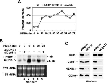
P-TEFb phosphorylates RNA polymerase II and negative elongation factors to stimulate general transcriptional elongation. It is kept in a functional equilibrium through alternately interacting with its positive (the Brd4 protein) and negative (the HEXIM1 protein and 7SK snRNA) regulators. To investigate the physiological significance of this phenomenon, we analyzed the responses of HeLa cells and murine erythroleukemia cells (MELC) to hexamethylene bisacetamide (HMBA), which inhibits growth and induces differentiation of many cell types. For both cell types, an efficient, albeit temporary disruption of the 7SK-HEXIM1-P-TEFb snRNP and enhanced formation of the Brd4-P-TEFb complex occurred soon after the treatment started. When the P-TEFb-dependent HEXIM1 expression markedly increased as the treatment continued, the abundant HEXIM1 pushed the P-TEFb equilibrium back toward the 7SK/HEXIM1-bound state. For HeLa cells, as HMBA produced only a minor, temporary effect on their growth, the equilibrium gradually returned to its pretreatment level. In contrast, long-term treatment of MELC induced terminal division and differentiation. Concurrently, the P-TEFb equilibrium was shifted overwhelmingly toward the 7SK snRNP side. Together, these data link the P-TEFb equilibrium to the intracellular transcriptional demand and proliferative/differentiated states of cells.


Figures

FIG. 1.
FIG. 1.
HMBA stimulates transcription from the HIV-1 promoter. (A) HMBA induces expression of the luciferase reporter gene driven by the HIV-1 LTR. A stable HeLa-based cell line containing an integrated HIV-1 LTR-luciferase reporter construct was treated with 10 mM HMBA for the indicated time periods. The level of induction (n-fold) in luciferase activity compared to that in untreated cells is shown. (B) The HMBA-induced increase in luciferase activity occurs at the mRNA level. Semiquantitative RT-PCR and Northern blot analyses were performed to detect luciferase mRNA from among total RNA isolated at various time points of a continuous HMBA treatment. The levels of the GAPDH mRNA and 7SK snRNA were also analyzed as internal controls. (C) NE from HMBA-treated HeLa cells significantly increases HIV-1 transcription in vitro. Reaction mixtures contained the transcription template HIV+TAR-G400 and NE prepared from cells treated with HMBA for 0 or 3 h. RNA fragments transcribed from a G-less cassette inserted into the template at a position ∼1 kb downstream of the HIV-1 promoter are indicated.
FIG. 2.
FIG. 2.
HMBA treatment of HeLa cells disrupts the 7SK snRNP and enhances the binding of P-TEFb to the HIV-1 chromatin template. (A) Treatment of HeLa cells with HMBA induces the dissociation of HEXIM1 and 7SK from P-TEFb. F1C2, a HeLa-based cell line stably expressing CDK9-f, was treated with HMBA for the indicated numbers of hours. CDK9-f, CycT1, HEXIM1, and 7SK present in NEs (lanes 1 to 5) and the anti-FLAG immunoprecipitates (αFLAG IP) (lanes 6 to 10) were detected by Western and Northern blotting. The presence of endogenous CDK9 (endo. CDK9) in NEs was revealed by anti-CDK9 Western blotting. (B) HMBA enhances the binding of P-TEFb to the HIV-1 chromatin template. The HeLa-based cell line with the integrated luciferase reporter gene driven by the HIV-1 LTR was treated with HMBA for 0 or 3 h. ChIP with anti-CDK9 antibody was performed. Three regions corresponding to the promoter region, interior region, and 3′ untranslated region (3′ UTR) of the integrated HIV-1 LTR-luciferase gene, as well as an interior region of the endogenous GAPDH gene, were PCR amplified from the precipitated and purified DNA. Numbers in parentheses indicate nucleotides. Amplified signals from 10% of the input chromatin are also shown.
FIG. 3.
FIG. 3.
Transient disruption of the 7SK snRNP, formation of the Brd4-P-TEFb complex, and activation of HIV-1 transcription in HMBA-treated HeLa cells. (A) Prolonged treatment with HMBA leads to reformation of the 7SK snRNP. HeLa cells were incubated with HMBA for the indicated time periods. Levels of HEXIM1 associated with the immunoprecipitated CDK9 derived from NEs were detected by Western blotting, quantified, and normalized to CDK9 levels, and they are shown as percentages relative to the pretreatment level, which was set to 100%. (B) HMBA transiently induces luciferase mRNA synthesis. Results are from Northern blot analysis of luciferase mRNA transcribed from the HIV-1 LTR in HeLa cells treated with HMBA for the indicated time periods. As a loading control, 7SK snRNA in total RNA samples was also examined. (C) Transient induction of luciferase activity in HMBA-treated cells. The level of induction (n-fold) in luciferase activity, expressed from the integrated HIV-1 LTR-luciferase reporter gene, was measured with a HeLa-based cell line treated with HMBA for the indicated time periods. pHIV, HIV-1 promoter. (D) P-TEFb alternately interacts with HEXIM1 and Brd4 throughout the course of HMBA treatment. NEs were prepared from HeLa cells treated with HMBA for the indicated durations and subjected to immunoprecipitation with anti-CDK9 antibody (αCDK9) (lanes 5 to 7) or, as a negative control (cntl.), anti-CDK4 antibody (lane 4). Brd4, HEXIM1, CycT1, and CDK9 present in the immunoprecipitates (IP) and NEs were examined by Western blotting.
FIG. 4.
FIG. 4.
P-TEFb-dependent induction of HEXIM1 expression by HMBA. (A) HMBA treatment of HeLa cells increases HEXIM1 expression. HEXIM1 levels in NE of HMBA-treated cells were detected by Western blotting at various time points of the treatment. The HEXIM1 signals were quantified and normalized to those of CDK9 for each time point, and they are shown as percentages relative to the pretreatment level, which was set to 100%. (B) P-TEFb is required for HMBA-induced HEXIM1 mRNA synthesis. HeLa cells transfected with either the empty pSuper vector (−) or vectors expressing the CDK9- and CycT1-specific siRNAs (siCDK9 and siCycT1, respectively) were treated with HMBA for 0, 6, or 24 h. Total RNA was isolated and subjected to Northern blot analysis to detect the major 2.5-kb and minor 4.0-kb HEXIM1 mRNA. The 18S and 28S rRNA present in total RNA were stained with ethidium bromide and used as loading controls. (C) CycT1- and Brd4-dependent production of the HEXIM1 protein. Western blotting was performed to examine the levels of Brd4, CycT1, HEXIM1, and CDK9 in NEs prepared from HeLa cells transfected with either the empty pSuper vector or the indicated siRNA-expressing pSuper constructs at 48 h posttransfection.
FIG. 5.
FIG. 5.
HeLa cells strive to maintain a constant level of nuclear 7SK snRNP for optimal growth. (A) HMBA suppresses HeLa cell growth only mildly and temporarily. Cells were either untreated or treated with HMBA for 6 or 24 h. After removal of the drug, cells were placed in fresh media at low but equal concentrations and allowed to grow for 2 or 4 days. Cell counts were determined and are shown as percentages relative to those of untreated cells, which were set to 100%. (B) Diversion of free HEXIM1 into the 7SK snRNP in HeLa cells expressing the HEXIM1-specific siRNAs. NEs were prepared from HeLa-based cell lines either containing the empty pSuper vector or expressing the indicated HEXIM1-specific siRNAs and examined for their HEXIM1 and CDK9 levels by Western blotting (lanes 1 to 5). Two independent cell clones (denoted by -1 and -2 after the siRNA designations) for each siRNA were analyzed. The NEs were also subjected to immunoprecipitation with either anti-CDK4 (αCDK4) (lane 6) or anti-CDK9 (lanes 7 to 11) antibody, and the indicated factors present in the immunoprecipitates (IP) were examined by Northern and Western blotting.
FIG. 6.
FIG. 6.
Enhanced sequestration of P-TEFb into the 7SK snRNP and elevated expression of HEXIM1 in HMBA-treated MELC. (A) HMBA treatment of MELC severely inhibits cell growth. HeLa cells and MELC were either untreated or treated with HMBA for 72 h. Upon removal of the drug, cells were placed in fresh media at low but equal concentrations and allowed to grow for 2 days. Cell counts were determined and are shown as percentages relative to those of untreated cells, which were set to 100%. (B) NEs were prepared from MELC treated with HMBA for the indicated time periods and subjected to anti-CDK9 (αCDK9) immunoprecipitation. HEXIM1 associated with the immunoprecipitates (IP) or present in NEs were detected by Western blotting. The HEXIM1 signals were normalized to those of CDK9 for each time point and are shown as percentages relative to the pretreatment levels, which were set to 100%. (C) Diagram depicting the regulation of P-TEFb activity during the course of HMBA treatment. The 7SK snRNP is proposed to contain two copies of P-TEFb and HEXIM1 and one copy of 7SK (14). During the first several hours of HMBA treatment, the 7SK snRNP was converted into the Brd4-P-TEFb complex, which stimulated P-TEFb-dependent HEXIM1 gene expression. The elevated HEXIM1 levels then pushed the P-TEFb equilibrium back toward the 7SK snRNP direction during a prolonged HMBA treatment. For details, see Discussion.

Similar articles

Cited by

References

    1. Barboric, M., and B. M. Peterlin. 2005. A new paradigm in eukaryotic biology: HIV Tat and the control of transcriptional elongation. PLoS Biol. 3:e76. - PMC - PubMed
    1. Bellan, C., G. De Falco, S. Lazzi, P. Micheli, S. Vicidomini, K. Schurfeld, T. Amato, A. Palumbo, L. Bagella, E. Sabattini, S. Bartolommei, M. Hummel, S. Pileri, P. Tosi, L. Leoncini, and A. Giordano. 2004. CDK9/CYCLIN T1 expression during normal lymphoid differentiation and malignant transformation. J. Pathol. 203:946-952. - PubMed
    1. Byers, S. A., J. P. Price, J. J. Cooper, Q. Li, and D. H. Price. 2005. HEXIM2, a HEXIM1-related protein, regulates positive transcription elongation factor b through association with 7SK. J. Biol. Chem. 280:16360-16367. - PubMed
    1. Chao, S.-H., and D. H. Price. 2001. Flavopiridol inactivates P-TEFb and blocks most RNA polymerase II transcription in vivo. J. Biol. Chem. 276:31793-31799. - PubMed
    1. Chen, R., Z. Yang, and Q. Zhou. 2004. Phosphorylated positive transcription elongation factor b (P-TEFb) is tagged for inhibition through association with 7SK snRNA. J. Biol. Chem. 279:4153-4160. - PubMed

Publication types

MeSH terms

LinkOut - more resources