. 2006 Oct;26(19):7167-77.
doi: 10.1128/MCB.00810-06.

Hematopoietic precursor cells transiently reestablish permissiveness for X inactivation

Affiliations

Hematopoietic precursor cells transiently reestablish permissiveness for X inactivation

Fabio Savarese et al. Mol Cell Biol. 2006 Oct.

Abstract

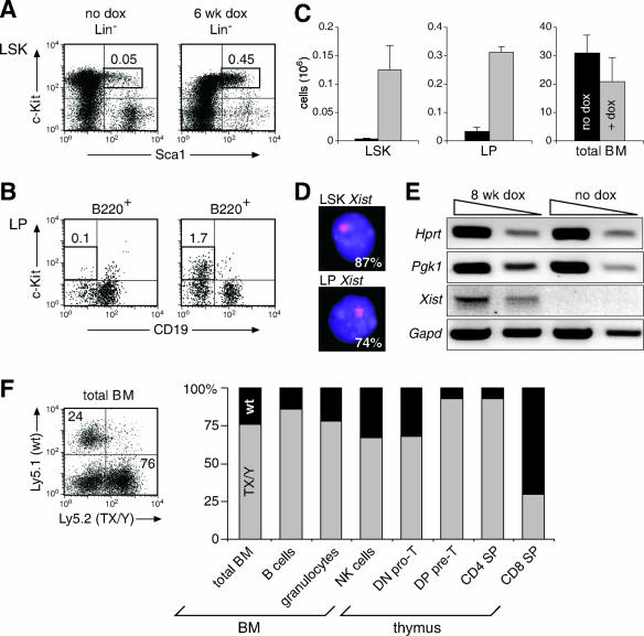
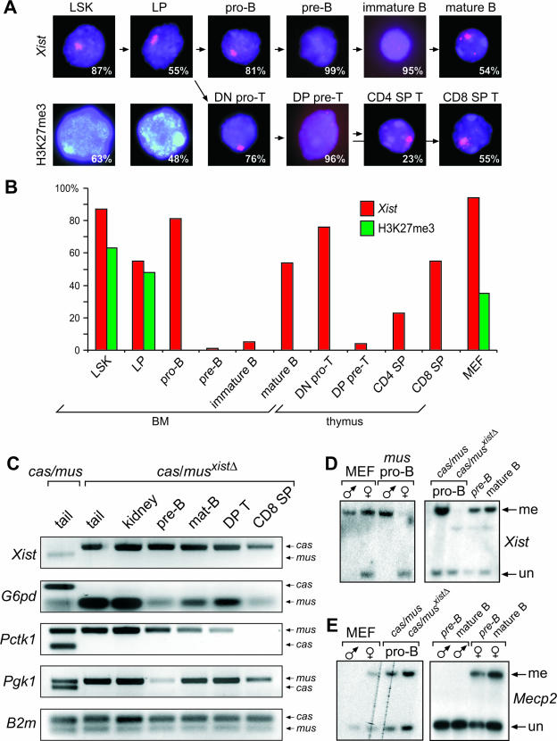
Xist is the trigger for X inactivation in female mammals. The long noncoding Xist RNA localizes along one of the two female X chromosomes and initiates chromosome-wide silencing in the early embryo. In differentiated cells, Xist becomes dispensable for the maintenance of the inactive X, and its function for initiation of silencing is lost. How Xist mediates gene repression remains an open question. Here, we use an inducible Xist allele in adult mice to identify cells in which Xist can cause chromosome-wide silencing. We show that Xist has the ability to initiate silencing in immature hematopoietic precursor cells. In contrast, hematopoietic stem cells and mature blood cells are unable to initiate ectopic X inactivation. This indicates that pathways critical for silencing are transiently activated in hematopoietic differentiation. Xist-responsive cell types in normal female mice show a change of chromatin marks on the inactive X. However, dosage compensation is maintained throughout hematopoiesis. Therefore, Xist can initiate silencing in precursors with concomitant maintenance of dosage compensation. This suggests that Xist function is restricted in development by the limited activity of epigenetic pathways rather than by a change in the responsiveness of chromatin between embryonic and differentiated cell types.


Figures

FIG. 1.
FIG. 1.
Ectopic X inactivation upon Xist induction in the embryo. (A) Generation of the inducible TX allele. The tet operator sequence (green) was introduced into the SacII site upstream of the P1 promoter of Xist. The probe XB1K is indicated. (B) Doxycycline (dox)-induced DNA binding of the transactivator protein nls-rtTA results in Xist expression. (C and D) Northern blot analysis of X-linked (C) and erythroid cell-specific (D) gene transcripts in male TX/Y R26rtTA/rtTA embryos, in which Xist was induced with doxycycline (+) for 4 days starting at the indicated time point. (E) Lateral view of TX/Y R26rtTA/rtTA embryos (+) after Xist induction for 4 days after the time point shown. Untreated (−) embryos were used as control.
FIG. 2.
FIG. 2.
Xist induction leads to anemia in adult mice. (A) Survival of TX/Y R26rtTA/rtTA and control X/Y R26rtTA/rtTA males (n = 15 each) after doxycycline treatment for 10 weeks. (B) Hematocrit levels of male TX/Y R26rtTA/rtTA and control X/Y R26rtTA/rtTA mice (n = 15 each) after doxycycline treatment for 3 to 6 weeks. (C) Hypocellularity of the bone marrow and absence of the thymus (thy) in a TX/Y R26rtTA/rtTA male after 6 weeks of doxycycline treatment (+). The thymus of an untreated (−) mouse is indicated by a dashed line.
FIG. 3.
FIG. 3.
Depletion of immature hematopoietic cell types by ectopic X inactivation. FACS analysis of the bone marrow (A to E) and thymus (F) of male TX/Y R26rtTA/rtTA mice without and with doxycycline treatment for 5 and 7 days (d). Pro-B cells (A), pre-B cells (B), immature and mature recirculating B cells (C), granulocytes and macrophages (D), erythroblasts (E), and DN, DP, and SP thymocytes (F) are shown with their percentage in the respective quadrant. Five mice per time point were analyzed to determine the absolute cell number (106) of the indicated cell types in the bone marrow (G) and thymus (H). (I) Northern blot analysis of in vitro cultured pro-B cells and ex vivo sorted pre-B cells and splenic IgD+ B cells before (−) or after (+) 4 days of doxycycline treatment. (K) Xist RNA FISH and H3K27me3 staining in 4-day-induced pre-B cells of a male TX/Y R26rtTA/rtTA mouse. The percentage of cells (n > 100) containing a signal is indicated. DNA is stained by DAPI (blue). BM, bone marrow; I, immature; R, recirculating; G, granulocytes; M, macrophages.
FIG. 4.
FIG. 4.
Absence of ectopic X inactivation in HSCs. (A to E) Male TX/Y R26rtTA/rtTA mice were induced with doxycycline for 6 weeks or left untreated. FACS analysis of multipotent LSK (A) and LPs (B). The percentage of total bone marrow cells in the respective gate is indicated. (C) Absolute cell numbers (106) of LSK, LPs, and total bone marrow cells (n = 5 each). (D) Xist RNA FISH signals with their detection frequency are shown for FACS-sorted LSK and LPs. DAPI staining is shown in blue. (E) RT-PCR analysis of X-linked gene expression in sorted LSK cells. Tenfold cDNA dilutions were analyzed. (F) FACS analysis of Ly5.1+ recipient mice 6 months after transplantation of a 1:1 mixture of bone marrow from a wild-type (wt) Ly5.1+ mouse and a TX/Y R26rtTA/rtTA Ly5.2+ mouse that was treated for 6 weeks with doxycycline. The relative contributions of wild-type (wt) Ly5.1+ HSCs (black bars) and TX/Y Ly5.2+ HSCs (gray bars) to the different lineages are shown. BM, bone marrow; dox, doxycycline.
FIG. 5.
FIG. 5.
Absence of markers of the Xi in immature lymphocytes of female mice. (A) Xist RNA FISH (red) and H3K27me3 staining (green) in hematopoietic cell types (n = 200 each) of a wild-type female mouse. The percentage of cells is indicated. DNA is stained by DAPI (blue). (B) Statistical evaluation of Xist and H3K27me3 signals in hematopoietic cells and mouse embryo fibroblasts. (C) X-linked G6pd, Pgk1, Pctk1, Xist, and autosomal control B2m expression was analyzed by allele-specific RT-PCR in the tissues indicated from an F1 cas/mus(xistΔ) female mouse, containing a maternal M. musculus X bearing a deletion of the Xist gene and a paternal M. castaneus X, and from a control wild-type cas/mus female. (D and E) Methylation analysis of the Xist (D) and Mecp2 promoter (E) in the indicated cell types. Mouse embryo fibroblasts and pro-B cells of all indicated sexes and genotypes were cultured, while pre-B and mature B cells were sorted ex vivo. MEF, mouse embryo fibroblast.

References

    1. Bacher, C. P., M. Guggiari, B. Brors, S. Augui, P. Clerc, P. Avner, R. Eils, and E. Heard. 2006. Transient colocalization of X-inactivation centres accompanies the initiation of X inactivation. Nat. Cell. Biol. - PubMed
    1. Balciunaite, G., R. Ceredig, S. Massa, and A. G. Rolink. 2005. A B220+ CD117+ CD19− hematopoietic progenitor with potent lymphoid andmyeloid developmental potential. Eur. J. Immunol. 2019-2030. - PubMed
    1. Bassing, C. H., W. Swat, and F. W. Alt. 2002. The mechanism and regulation of chromosomal V(D)J recombination. Cell 109(Suppl.):S45-S55. - PubMed
    1. Borsani, G., R. Tonlorenzi, M. C. Simmler, L. Dandolo, D. Arnaud, V. Capra, M. Grompe, A. Pizzuti, D. Muzny, C. Lawrence, et al. 1991. Characterization of a murine gene expressed from the inactive X chromosome. Nature 351:325-329. - PubMed
    1. Brockdorff, N. 2002. X-chromosome inactivation: closing in on proteins that bind Xist RNA. Trends Genet. 18:352-358. - PubMed

Publication types

MeSH terms