Comparative Study
. 2006 Oct;26(19):7299-317.
doi: 10.1128/MCB.02405-05.

Bioinformatic and comparative localization of Rab proteins reveals functional insights into the uncharacterized GTPases Ypt10p and Ypt11p

Affiliations
Comparative Study

Bioinformatic and comparative localization of Rab proteins reveals functional insights into the uncharacterized GTPases Ypt10p and Ypt11p

Stéphanie Buvelot Frei et al. Mol Cell Biol. 2006 Oct.

Abstract

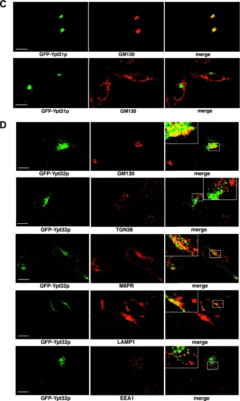
A striking characteristic of a Rab protein is its steady-state localization to the cytosolic surface of a particular subcellular membrane. In this study, we have undertaken a combined bioinformatic and experimental approach to examine the evolutionary conservation of Rab protein localization. A comprehensive primary sequence classification shows that 10 out of the 11 Rab proteins identified in the yeast (Saccharomyces cerevisiae) genome can be grouped within a major subclass, each comprising multiple Rab orthologs from diverse species. We compared the locations of individual yeast Rab proteins with their localizations following ectopic expression in mammalian cells. Our results suggest that green fluorescent protein-tagged Rab proteins maintain localizations across large evolutionary distances and that the major known player in the Rab localization pathway, mammalian Rab-GDI, is able to function in yeast. These findings enable us to provide insight into novel gene functions and classify the uncharacterized Rab proteins Ypt10p (YBR264C) as being involved in endocytic function and Ypt11p (YNL304W) as being localized to the endoplasmic reticulum, where we demonstrate it is required for organelle inheritance.


Figures

FIG. 1.
FIG. 1.
Global view of Rab sequence space with two-dimensional principal components analysis. (A) The x and y axes represent the values of the second and third principal components, respectively. The analysis was performed on a database containing 560 individually checked and unique Rab sequences, including each Rab protein identified in S. cerevisiae. Automatic clustering with the Clusterdata function in Matlab was performed to identify major groupings in the data. This analysis identified 10 groups which are color coded and named according to a representative mammalian member of the group. (B) The position of each Rab protein present in the S. cerevisiae genome is indicated in relation to the global Rab sequence space. For a list of the Rab proteins in yeast and accession numbers, see the supplemental material.
FIG. 2.
FIG. 2.
Functionality of GFP-tagged constructs. (A) GFP-tagged SEC4 and YPT1 constructs were transformed into SEC4Δ and YPT1Δ tester strains and streaked onto medium with (+) or without (−) 5-FOA at 25°C to assess functionality. This assay was performed in comparison to yeast transformed with empty vector as a negative control and wild-type SEC4 and YPT1 as positive controls. (B) ypt6Δ cells were assayed for survival at 37°C when transformed with empty vector-, YPT6-, or GFP-YPT6-containing plasmids. (C) ypt31Δ ypt31ts cells were assayed for the ability of GFP-tagged YPT31 to rescue growth at 37°C compared to that of vector alone. (D) vps21Δ cells were assayed for the uptake of lucifer yellow CH into the vacuole in the presence (+) or absence (−) of GFP-Vps21p. (E) ypt7Δ cells with GFP-YPT7 (i), vector only (ii), and wild-type YPT7 (iii) constructs were assayed for vacuolar morphologies with FM4-64. DIC, differential interference contrast. (F) ypt10Δ cells with GFP-YPT10 (i), vector only (ii), and wild-type YPT10 (iii) expressed behind the copper-inducible promoter PCUP1 were assayed for growth on media ± 0.7 mM CuSO4.
FIG. 3.
FIG. 3.
Localization of GFP-Ypt6p in yeast and HeLa cells. (A) GFP-Ypt6p was expressed in yeast cells, and live cells were viewed by epifluorescence microscopy. The GFP fluorescence signal is presented with differential interference contrast (DIC) images of the same cells. (B) GFP-Ypt6p was expressed in HeLa cells, and fixed cells were viewed by confocal microscopy. The GFP fluorescence signal is compared to those of antibody markers for the following cellular compartments: (i) Golgi apparatus GM130, (ii) trans-Golgi network TGN38, and (iii and iv) transferrin internalized for 15 min. In each case, a merge of the two fluorescence images is shown. Bars = 10 μm.
FIG.4.
FIG.4.
Localization of GFP-Vps21p, GFP-Ypt52p, and GFP-Ypt53p in yeast and HeLa cells. (A) Localization of GFP-Vps21p, GFP-Ypt52p, and GFP-Ypt53p in yeast. (i) GFP-Vps21p was expressed in yeast cells, and live cells were viewed by epifluorescence microscopy. The GFP fluorescence signal is presented with differential interference contrast images of the same cells. A merge of the two images is shown. (ii) GFP-Ypt52p-expressing yeast cells. Conditions were as described for panel i. (iii) GFP-Ypt53p-expressing yeast cells. Conditions were as described for panel i. (B) Localization of GFP-Ypt51p in HeLa cells. GFP-Vps21p was expressed in HeLa cells that were fixed and viewed by confocal microscopy. The GFP fluorescence signal was compared to those of antibody markers for the following cellular compartments: early endosomes (EEA1), late endosomes (M6PR), and late endosomes/lysosomes (LAMP1). In each case, a merge of the two fluorescence images is shown. Insets show selected areas enlarged approximately threefold, with color levels optimized to show colocalization. Bars = 10 μm. (C) Localization of GFP-Ypt52p in HeLa cells. GFP-Ypt52p was expressed in HeLa cells, and fixed cells were viewed by confocal microscopy. The GFP fluorescence signal was compared to those of antibody markers for the following cellular compartments: early endosomes (EEA1), late endosomes (M6PR), and late endosomes/lysosomes (LAMP1). In each case, a merge of the two fluorescence images is shown. Insets show selected areas enlarged approximately threefold, with color levels optimized to show colocalization. Bars = 10 μm. (D) Localization of GFP-Ypt53p in HeLa cells. GFP-Ypt53p was expressed in HeLa cells, and fixed cells were viewed by confocal microscopy. The GFP fluorescence signal was compared to those of antibody markers for the following cellular compartments: early endosomes (EEA1), late endosomes (M6PR), and late endosomes/lysosomes (LAMP1). In each case, a merge of the two fluorescence images is shown. Insets show selected areas enlarged approximately threefold, with color levels optimized to show colocalization. Bars = 10 μm.
FIG.4.
FIG.4.
Localization of GFP-Vps21p, GFP-Ypt52p, and GFP-Ypt53p in yeast and HeLa cells. (A) Localization of GFP-Vps21p, GFP-Ypt52p, and GFP-Ypt53p in yeast. (i) GFP-Vps21p was expressed in yeast cells, and live cells were viewed by epifluorescence microscopy. The GFP fluorescence signal is presented with differential interference contrast images of the same cells. A merge of the two images is shown. (ii) GFP-Ypt52p-expressing yeast cells. Conditions were as described for panel i. (iii) GFP-Ypt53p-expressing yeast cells. Conditions were as described for panel i. (B) Localization of GFP-Ypt51p in HeLa cells. GFP-Vps21p was expressed in HeLa cells that were fixed and viewed by confocal microscopy. The GFP fluorescence signal was compared to those of antibody markers for the following cellular compartments: early endosomes (EEA1), late endosomes (M6PR), and late endosomes/lysosomes (LAMP1). In each case, a merge of the two fluorescence images is shown. Insets show selected areas enlarged approximately threefold, with color levels optimized to show colocalization. Bars = 10 μm. (C) Localization of GFP-Ypt52p in HeLa cells. GFP-Ypt52p was expressed in HeLa cells, and fixed cells were viewed by confocal microscopy. The GFP fluorescence signal was compared to those of antibody markers for the following cellular compartments: early endosomes (EEA1), late endosomes (M6PR), and late endosomes/lysosomes (LAMP1). In each case, a merge of the two fluorescence images is shown. Insets show selected areas enlarged approximately threefold, with color levels optimized to show colocalization. Bars = 10 μm. (D) Localization of GFP-Ypt53p in HeLa cells. GFP-Ypt53p was expressed in HeLa cells, and fixed cells were viewed by confocal microscopy. The GFP fluorescence signal was compared to those of antibody markers for the following cellular compartments: early endosomes (EEA1), late endosomes (M6PR), and late endosomes/lysosomes (LAMP1). In each case, a merge of the two fluorescence images is shown. Insets show selected areas enlarged approximately threefold, with color levels optimized to show colocalization. Bars = 10 μm.
FIG. 5.
FIG. 5.
Localization of GFP-Ypt31p and GFP-Ypt32p in yeast and HeLa cells. (A) Localization of GFP-Ypt31p in yeast. GFP-Ypt31p-expressing live cells were viewed by fluorescence microscopy. GFP-Ypt31p localization was analyzed at various stages of the yeast cell cycle. A nuclear marker (Gal4BD-RFP) and the relative sizes of the mother and daughter cells were used to ascertain the cell cycle stage. The overlay of the GFP and RFP channels are of the maximum projection of each channel, and differential interference contrast (DIC) images were taken in one z plane. (B) Localization of GFP-Ypt32p in yeast. GFP-Ypt32p was expressed in yeast cells as the only copy, and live cells were viewed by fluorescence microscopy as described for panel A. (C) Localization of GFP-Ypt31p in HeLa cells. GFP-Ypt31p was expressed in HeLa cells, and fixed cells were viewed by confocal microscopy. The GFP fluorescence signal was compared to those of antibody markers for the following cellular compartments: Golgi apparatus (GM130) and trans-Golgi network (TGN38). In each case, a merge of the two fluorescence images is shown. Bars = 10 μm. (D) Localization of GFP-Ypt32p in HeLa cells. GFP-Ypt32p was expressed in HeLa cells, and fixed cells were viewed by confocal microscopy. The GFP fluorescence signal was compared to those of antibody markers for the following cellular compartments: Golgi apparatus (GM130) and trans-Golgi network (TGN38), late endosomes (M6PR), late endosomes/lysosomes (LAMP1), and early endosomes (EEA1). In each case, a merge of the two fluorescence images is shown. Insets show selected areas enlarged approximately threefold, with color levels optimized to show colocalization. Bars = 10 μm.
FIG. 5.
FIG. 5.
Localization of GFP-Ypt31p and GFP-Ypt32p in yeast and HeLa cells. (A) Localization of GFP-Ypt31p in yeast. GFP-Ypt31p-expressing live cells were viewed by fluorescence microscopy. GFP-Ypt31p localization was analyzed at various stages of the yeast cell cycle. A nuclear marker (Gal4BD-RFP) and the relative sizes of the mother and daughter cells were used to ascertain the cell cycle stage. The overlay of the GFP and RFP channels are of the maximum projection of each channel, and differential interference contrast (DIC) images were taken in one z plane. (B) Localization of GFP-Ypt32p in yeast. GFP-Ypt32p was expressed in yeast cells as the only copy, and live cells were viewed by fluorescence microscopy as described for panel A. (C) Localization of GFP-Ypt31p in HeLa cells. GFP-Ypt31p was expressed in HeLa cells, and fixed cells were viewed by confocal microscopy. The GFP fluorescence signal was compared to those of antibody markers for the following cellular compartments: Golgi apparatus (GM130) and trans-Golgi network (TGN38). In each case, a merge of the two fluorescence images is shown. Bars = 10 μm. (D) Localization of GFP-Ypt32p in HeLa cells. GFP-Ypt32p was expressed in HeLa cells, and fixed cells were viewed by confocal microscopy. The GFP fluorescence signal was compared to those of antibody markers for the following cellular compartments: Golgi apparatus (GM130) and trans-Golgi network (TGN38), late endosomes (M6PR), late endosomes/lysosomes (LAMP1), and early endosomes (EEA1). In each case, a merge of the two fluorescence images is shown. Insets show selected areas enlarged approximately threefold, with color levels optimized to show colocalization. Bars = 10 μm.
FIG. 6.
FIG. 6.
Localization of GFP-Ypt10p in yeast and HeLa cells. (A) GFP-Ypt10p was expressed in yeast cells, live cells were viewed by epifluorescence microscopy, and fluorescence images were subjected to double-blind deconvolution. The GFP fluorescence signal is presented with a differential interference contrast (DIC) image of the same cells. A merge of the two images is shown. (i) GFP-Ypt10p expressed from endogenous promoter in ypt10Δ cells; (ii) GFP-Ypt10p expressed with PCUP1 (image shows basal expression of gene in absence of Cu3+ addition to media); (iii) live-cell imaging of GFP-Ypt10p in cells labeled with FM4-64 for 15 min followed by a 45-min washout to identify vacuolar membranes. (B) GFP-Ypt10p was expressed in HeLa cells, fixed and permeabilized, and labeled with TRITC-phalloidin to visualize the actin cytoskeleton. Cells were viewed by epifluorescence microscopy, and images were subjected to double-blind deconvolution. A single transverse slice though the cell is shown. GFP and TRITC-phalloidin are shown both individually and as a merged image. The panels below show GFP-Ypt10p in the periphery of the cell (i) and in conjunction with markers of the Golgi apparatus: GM130 (ii), GFP early endosome EEA1 (iii), and the recycling endosome for Rab11 (iv).For panels ii, iii, and iv, GFP and antibody markers are shown in separate images.
FIG. 7.
FIG. 7.
Localization of GFP-Ypt11p in yeast and HeLa cells. (A) GFP-Ypt11p was expressed in yeast cells, and live cells were stained with Hoechst 33258. Cells were viewed by epifluorescence microscopy, and fluorescence images were subjected to double-blind deconvolution. The GFP fluorescence signal is presented with a differential interference contrast (DIC) image of the same cells, and a merge of the three images is shown. (B) GFP-Ypt11p was expressed in yeast cells, fixed, and stained with Hoechst 33258. Cells were analyzed by immunofluorescence microscopy using anti-Pdi1p antibodies. The GFP and immunofluorescence signal is presented with a differential interference contrast (DIC) image of the same cells. (C) GFP-Ypt11p was expressed in BHK cells and viewed by epifluorescence microscopy, and fluorescence images were subjected to double-blind deconvolution. Both a maximum projection and a single slice through the cell are shown. (D) GFP-Ypt11p was expressed in BHK cells and live cells incubated with ER-Tracker Blue-White DPX. Cells were fixed and viewed by epifluorescence microscopy. The GFP and ER-Tracker DPX signals are shown individually, and in a merged image with ER-Tracker DPX false-colored red to show colocalization with GFP. (E) GFP-Ypt11p was expressed in HeLa cells, and fixed cells were viewed by confocal microscopy. The GFP fluorescence signal was compared to that of an antibody marker for the endoplasmic reticulum, PDI. Images show the periphery of the cell both individually and as a merge of the two fluorescence images. (F) GFP-Ypt11p was expressed in BHK cells, and fixed cells were viewed by confocal microscopy. The GFP fluorescence signal was compared to that of an antibody marker for the ERGIC, KDEL receptor (KDEL-R). GFP and the KDEL-R are shown both individually and as a merged image. In the lower panels, the cells were incubated for 3 h at 15°C before fixation.
FIG. 8.
FIG. 8.
ypt11Δ cells influence the inheritance of ER membrane markers to a similar extent as observed for myo4Δ cells. (A) Wild-type, myo4Δ, ypt11Δ, and myo4Δ ypt11Δ cells expressing Hmg1p-GFP at the endogenous locus were grown to mid-log phase at 30°C in rich medium and then resuspended in nonfluorescent medium. A z stack image was taken, with the plane going through the bud neck being kept for further investigation. Representative pictures are shown for each genotype. (B) Wild-type, myo4Δ, ypt11Δ, and myo4Δ ypt11Δ cells expressing Sec61p-GFP were photographed as described for panel A. Images were processed as for panel A, with a representative picture shown for each genotype. (C and D) Pictures shown in panel A (C) or B (D) were analyzed using Image J 1.29 software (http://rsb.info.nih.gov/ij). Four sets of data were extracted from the photographs: the intensity signal of the bud cortex, the intensity signal of the mother cortex, the area of the bud, and the area of the mother. Each dot represents one cell and is the ratio of the intensity of the bud cortex/intensity of the mother cortex over the area of the bud/the area of the mother. The experiment was repeated three times, and only one of the experiments is represented in the graph; the tendencies were similar in all three experiments. Table 1 shows a summary quantification of the data set.
FIG. 9.
FIG. 9.
Functional replacement of SEC19/GDI1 with mammalian Rab-GDI. (A) sec19-1 suppression. sec19-1 cells were transformed with the following plasmids: (1) empty vector, (2) mammalian Rab-GDI, (3) SEC19/GDI1-positive control, as indicated before testing for growth on the permissive and restrictive temperature on yeast-peptone medium containing 1.5% raffinose with 0.5% galactose. (B) Ability of mammalian Rab-GDI constructs to serve as the sole source of yeast SEC19/GDI1. GDI1Δ cells were transformed with constructs as indicated before testing for growth on 5-FOA-containing plates with either glucose or galactose as a carbon source. These constructs are indicated in the plate schematic.

References

    1. Ali, B. R., and M. C. Seabra. 2005. Targeting of Rab GTPases to cellular membranes. Biochem. Soc. Trans. 33:652-656. - PubMed
    1. Ali, B. R., C. Wasmeier, L. Lamoreux, M. Strom, and M. C. Seabra. 2004. Multiple regions contribute to membrane targeting of Rab GTPases. J. Cell Sci. 117:6401-6412. - PubMed
    1. Bacon, R. A., A. Salminen, H. Ruohola, P. Novick, and S. Ferro-Novick. 1989. The GTP-binding protein Ypt1 is required for transport in vitro: the Golgi apparatus is defective in ypt1 mutants. J. Cell Biol. 109:1015-1022. - PMC - PubMed
    1. Benli, M., F. Doring, D. G. Robinson, X. Yang, and D. Gallwitz. 1996. Two GTPase isoforms, Ypt31p and Ypt32p, are essential for Golgi function in yeast. EMBO J. 15:6460-6475. - PMC - PubMed
    1. Bereiter-Hahn, J. 1990. Behavior of mitochondria in the living cell. Int. Rev. Cytol. 122:1-63. - PubMed

Publication types

MeSH terms

Substances