Comparative Study
. 2006 Oct;9(10):1247-56.
doi: 10.1038/nn1775. Epub 2006 Sep 17.

Asymmetrical beta-actin mRNA translation in growth cones mediates attractive turning to netrin-1

Affiliations
Comparative Study

Asymmetrical beta-actin mRNA translation in growth cones mediates attractive turning to netrin-1

Kin-Mei Leung et al. Nat Neurosci. 2006 Oct.

Abstract

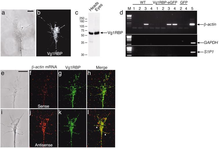
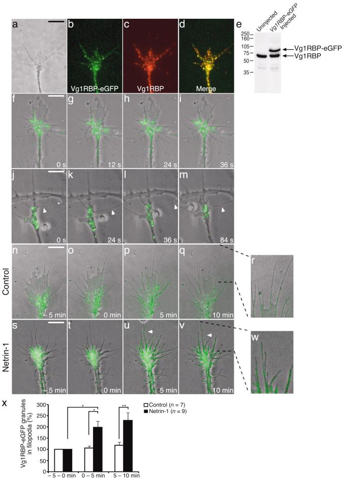
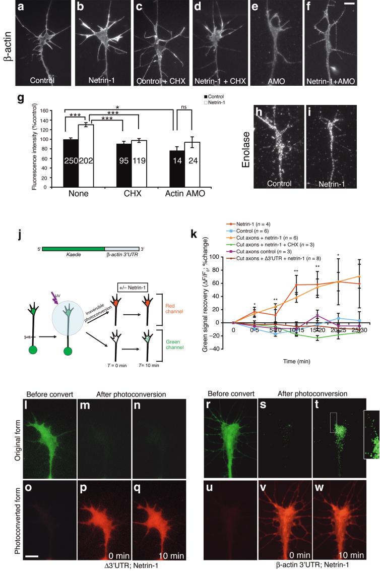
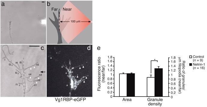
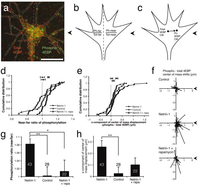
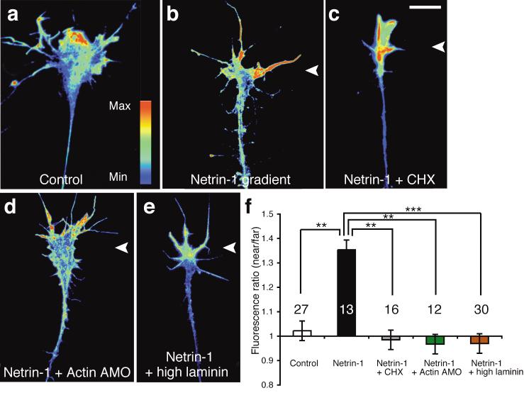
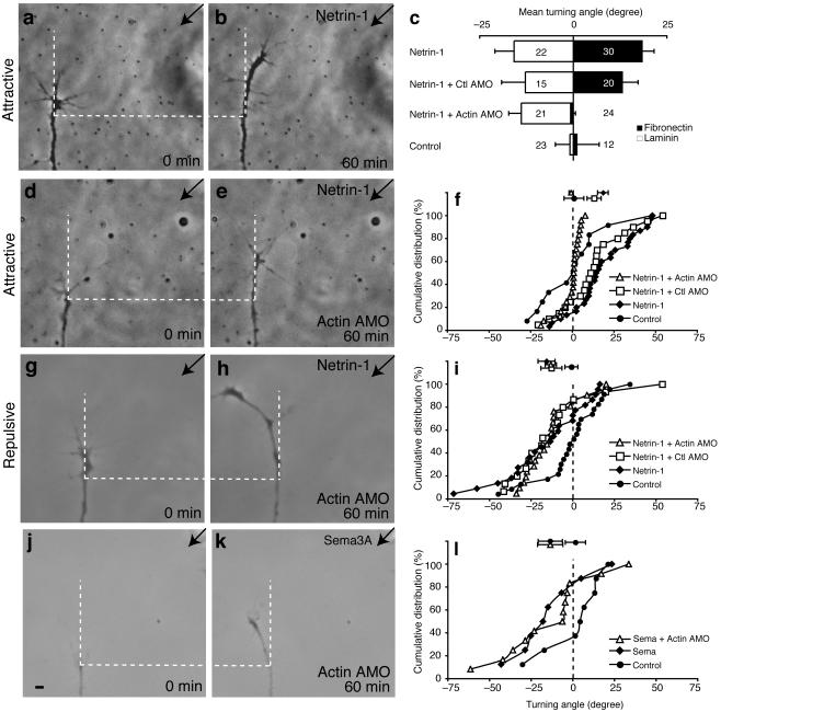
Local protein synthesis regulates the turning of growth cones to guidance cues, yet little is known about which proteins are synthesized or how they contribute to directional steering. Here we show that beta-actin mRNA resides in Xenopus laevis retinal growth cones where it binds to the RNA-binding protein Vg1RBP. Netrin-1 induces the movement of Vg1RBP granules into filopodia, suggesting that it may direct the localization and translation of mRNAs in growth cones. Indeed, a gradient of netrin-1 activates a translation initiation regulator, eIF-4E-binding protein 1 (4EBP), asymmetrically and triggers a polarized increase in beta-actin translation on the near side of the growth cone before growth cone turning. Inhibition of beta-actin translation abolishes both the asymmetric rise in beta-actin and attractive, but not repulsive, turning. Our data suggest that newly synthesized beta-actin, concentrated near sites of signal reception, provides the directional bias for polymerizing actin in the direction of an attractive stimulus.


Figures

Figure 1
Figure 1. Vg1RBP is expressed in retinal growth cones and interacts with β-actin mRNA.
Cultured stage 33/34 retinal explants were stained with Vg1RBP antibody (a: phase, b: anti-Vg1RBP). Vg1RBP is detected in the axon shaft, central domain and filopodia. Western blot analysis using Vg1RBP antibody shows a predominant 65 kDa band in stage 33/34 head or eye lysates (c). Vg1RBP was immunoprecipitated from head lysates of stage 33/34 wildtype (WT) embryos, injected with Vg1RBP-eGFP mRNA or GFP mRNA (d). RNA extracted from the immunoprecipitates was subjected to RT-PCR using primers to the 3’UTR of β-actin mRNA (top panel), GAPDH mRNA (middle panel) or S1P1 mRNA (bottom panel). 1: protein A beads only. 2: purified rabbit IgG. 3: Vg1RBP antiserum. 4: GFP antiserum. 5: Positive RT-PCR control. M: molecular DNA marker. β-actin mRNA is immunoprecipitated with both Vg1RBP and Vg1RBP-eGFP. GAPDH and S1P1 mRNAs do not interact with Vg1RBP. FISH using riboprobes directed against 3’UTR of β-actin mRNA (j, antisense) or control RNA (f, sense) together with immunostaining of Vg1RBP (g,k) reveals partial colocalization of Vg1RBP and β-actin mRNA in the growth cone (e,i: phase, f: control sense RNA, j: β-actin antisense RNA, g,k: Vg1RBP, h,l: merge). Arrowheads refer to complexes of Vg1RBP and β-actin mRNA in a filopodium. Scale bars 5 μm.
Figure 2
Figure 2. Netrin-1 or cell-contact induce translocation of Vg1RBP-eGFP into filopodia.
Retinal explants taken from stage 33/34 embryos expressingVg1RBP-eGFP were stained with Vg1RBP antibody. Both endogenous and overexpressed proteins are recognized by the Vg1RBP antibody and show similar patterns of localization (a: phase, b: Vg1RBP-eGFP, c: Vg1RBP, d: merge). Vg1RBP-eGFP is expressed at similar levels as the endogenous Vg1RBP as shown by Western blot analysis of head lysates from injected and uninjected embryos (e). Time-lapse analysis of Vg1RBP-eGFP shows bidirectional movement of Vg1RBP-eGFP granules in the axon shaft, growth cone central domain and filopodia. Images were taken every 12 seconds (f-i and Supplementary movie 1). Vg1RBP-eGFP granules are also detected moving along filopodial contact-contact sites (j-m, arrowheads and Supplementary movie 2). The asterisk indicates the branch/filopodium, which is going to contact the Vg1RBP-eGFP expressing filopodium. Live imaging of Vg1RBP-eGFP expressing retinal growth cones stimulated with control medium (n-r) or netrin-1 (s-w and Supplementary movie 3) at T = 0 min. The percentage of Vg1RBP-eGFP granules in the filopodia was calculated in each frame and averaged per 5 minutes (x). Application of netrin-1 results in an increase of Vg1RBP-eGFP granules in the filopodia compared to control. * P < 0.05, ** P < 0.01, Kruskal-Wallis test. Error bars s.e.m. Scale bars 5 μm.
Figure 3
Figure 3. Netrin-1 induces β-actin translation driven by its 3’UTR.
Stage 24 retinal growth cones were stimulated with netrin-1 for 5 minutes, stained for β-actin and fluorescence intensities were measured. Netrin-1induced an increase in β-actin QIF signal, which was blocked by CHX and β-actin AMO (a-g). *** P < 0.0001, Kruskal-Wallis test. Growth cones from β-actin AMO injected retina showed reduced β-actin QIF signal (e,g), which was not affected by netrin-1 stimulation (f,g). * P = 0.02, ns = non-significant, Mann-Whitney test. Enolase QIF signal was not affected by netrin-1 stimulation (h,i). Schematic diagram of Kaede construct and experimental design (j). Time-lapse imaging showed that only Kaede-green was detected before photoconversion (l,r). At T = 0 min, Kaede was photoconverted and only Kaede-red was detected (p,v). Netrin-1 had no effect on Kaede-green (l-n) or Kaede-red signals (o-q) in growth cones expressing Kaede-Δ3’UTR (k). However, netrin-1 induced a recovery of Kaede-green in both intact and severed growth cones expressing Kaede-β actin 3’UTR (k,s,t), which was blocked by CHX (k). The recovery was also seen in some filopodia (t, boxed area and inset). The exposure gain for inset picture is increased for visualization of filopodia. Kaede-red remained unchanged (v,w). The change in Kaede-green signal (ΔF) was compared to the image taken at T = 0 min (F0) and presented as ΔF/F0. * P < 0.05, ** P < 0.01, Mann-Whitney test. Scale bars 5 μm. Error bars s.e.m. Numbers inside bars indicate the number of growth cones analyzed.
Figure 4
Figure 4. Vg1RBP granules move into filopodia closest to a netrin-1 source.
Experimental design showing the pipette tip positioned 100 μm from the growth cone at an angle of 90°; a dashed line divides the growth cone into ‘near’ and ‘far’ sides with respect to the pipette. (a,b). Vg1RBP-eGFP expressing retinal growth cones were stimulated with a gradient of culture medium (control) or netrin-1 for 5 minutes and subsequently fixed (c-e). Mean fluorescence intensities as well as the number of granules per μm filopodia were calculated for both the near and far sides (c-e). A gradient of netrin-1 induces an increase in the number of Vg1RBP-eGFP granules per μm filopodia at the near side. No difference in Vg1RBP fluorescence intensity is observed in the far versus the near side (e). Near-side and far-side filopodia are numbered n1-n5 and f1-f5 respectively. Arrowheads indicate filopodia containing low-density (grey arrowheads) and high-density (white arrowheads) areas of Vg1RBP-eGFP granules. * P < 0.03 Mann Whitney test. Arrow indicates direction of pipette. Error bars s.e.m. Scale bars 5 μm.
Figure 5
Figure 5. Netrin-1 gradient causes asymmetric activation of translation regulator.
Growth cone double-stained for total-4EBP (red) and phospho-4EBP (green) with a line dividing near/far sides (a). Lack of overlap may be due to stochastic mutual exclusion by large Zenon Fc-antibody complexes (see Methods). Asymmetric phosphorylation of 4EBP was assessed by the near/far ratio method (b) or by the ‘center of mass’ method (c). Phosphorylation is higher on the near side than on the far side in netrin-1-stimulated growth cones. (P < 0.000001, paired t-test), but not control-stimulated growth cones (P = 0.9) or netrin-1-stimulated growth cones treated with rapamycin (d,g; P > 0.95). The center of mass of phospho-4EBP staining is significantly closer to the pipette than that of total-4EBP in netrin-1-stimulated growth cones (P < 0.0001, paired t-test), but not in control-stimulated growth cones (P > 0.65) or netrin-1-stimulated growth cones treated with rapamycin (e,h; P > 0.1). The difference in center of mass shifts between the netrin-1 and netrin-1 + rapamycin conditions is almost significant (P = 0.06, Welch-corrected unpaired t-test). Vector plots of center of mass shifts show that only the netrin-1 condition exhibits consistent center of mass shifts toward the pipette (f). Each vector represents the center of mass shift of phospho-4EBP relative to total-4EBP in one growth cone. The axon shaft is down. Numbers in (g) and (h) indicate number of growth cones per condition. ** P < 0.005, * P < 0.05, Welch-corrected unpaired t-test. Arrowheads indicate direction of pipette. Scale bar 10 μm. Error bars s.e.m.
Figure 6
Figure 6. Netrin-1 gradient elicits asymmetric increase of β-actin across the growth cone.
Growth cones from stage 24 retinal explants were exposed to a gradient of netrin-1 for 5 minutes and then stained for β-actin. Fluorescence intensities of both sides of the growth cone were measured and the near/far ratios of the different groups were compared (f). In control condition, β-actin was expressed at similar levels on both sides of the growth cone (a). Netrin-1 gradient induced an asymmetric rise in β-actin QIF signal on the near side (b), which was blocked by CHX (c) and β-actin AMO (d). Netrin-1 did not cause β-actin asymmetry in growth cones grown on high laminin substrate (e). Images were pseudo-coloured (see colour bar in a). ** P = 0.003, *** P < 0.001, Kruskal-Wallis test. Scale bar 10 μm. Error bars s.e.m. Numbers inside bars indicate the number of growth cones analyzed.
Figure 7
Figure 7. β-actin morpholinos block netrin-1-induced attractive turning.
Eye primordia from stage 24 embryos injected with β-actin or control AMO were cultured for 24 hours. Under attractive conditions, netrin-1 caused strong positive turning (a-c,f; 18.4° ± 3.2°) whereas control medium did not (0.9° ± 6.0°). This attractive response was blocked in β-actin AMO-containing growth cones (c-f; −0.7° ± 1.3°) whereas growth cones containing control AMO retained attractive turning in response to netrin-1 (c and f; 13.1° ± 4.2°). *** P < 0.001. Under repulsive conditions, growth cones turned away from the source of netrin-1 (c and i, −15.8° ± 5.1°) but not control medium (−0.9° ± 3.9°). Introduction of β-actin AMO, as well as control AMO, into growth cones did not affect the repulsive turning response (c,g-i; −13.2° ± 3.3° and −12.8° ± 6.3° respectively). *** P < 0.001. The repulsive turning triggered by Sema3A in control growth cones (−13.6° ±7.3°) is similar to that in β-actin AMO-containing growth cones (j-l; −13.6° ±7.5°). P > 0.05. The control supernatant did not cause repulsive turning (1.5° ± 6.1°). Kolmogorov-Smirnov test. Mean turning angle ± s.e.m.. Error bars s.e.m.

Comment in

  • Turning by asymmetric actin.
    Ming GL. Ming GL. Nat Neurosci. 2006 Oct;9(10):1201-3. doi: 10.1038/nn1006-1201. Nat Neurosci. 2006. PMID: 17001333 No abstract available.

Similar articles

Cited by

References

    1. Henley J, Poo MM. Guiding neuronal growth cones using Ca2+ signals. Trends Cell Biol. 2004;14:320–30. - PMC - PubMed
    1. Guirland C, Suzuki S, Kojima M, Lu B, Zheng JQ. Lipid rafts mediate chemotropic guidance of nerve growth cones. Neuron. 2004;42:51–62. - PubMed
    1. Wen Z, Guirland C, Ming GL, Zheng JQ. A CaMKII/calcineurin switch controls the direction of Ca(2+)-dependent growth cone guidance. Neuron. 2004;43:835–46. - PubMed
    1. Zheng JQ. Turning of nerve growth cones induced by localized increases in intracellular calcium ions. Nature. 2000;403:89–93. - PubMed
    1. Li Y, et al. Essential role of TRPC channels in the guidance of nerve growth cones by brain-derived neurotrophic factor. Nature. 2005;434:894–8. - PubMed

Publication types

MeSH terms