. 2006 Oct;116(10):2633-42.
doi: 10.1172/JCI28546. Epub 2006 Sep 14.

Selective tyrosine kinase inhibition by imatinib mesylate for the treatment of autoimmune arthritis

Affiliations

Selective tyrosine kinase inhibition by imatinib mesylate for the treatment of autoimmune arthritis

Ricardo T Paniagua et al. J Clin Invest. 2006 Oct.

Abstract

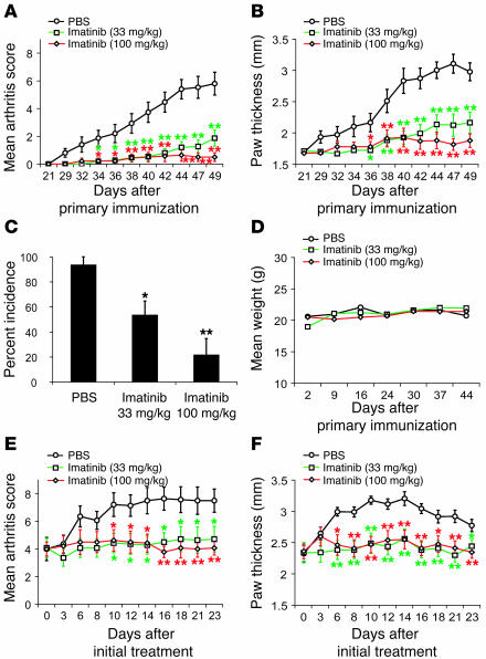
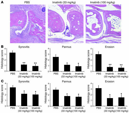
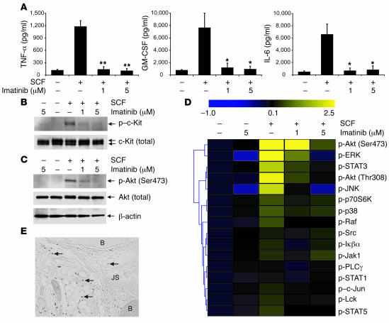
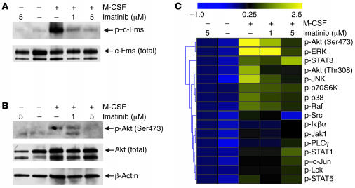
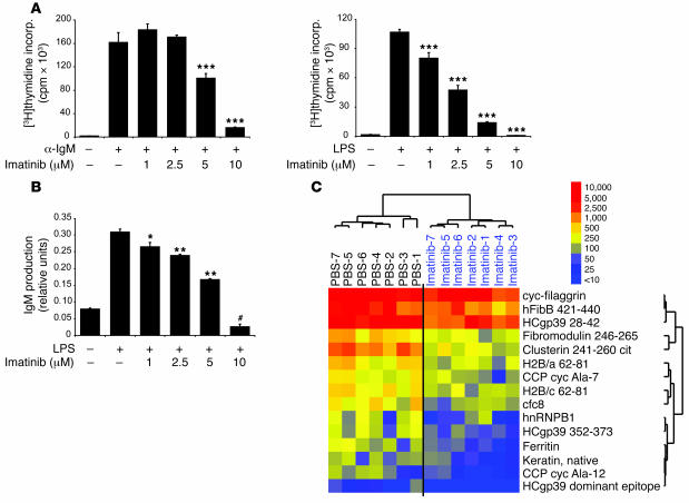
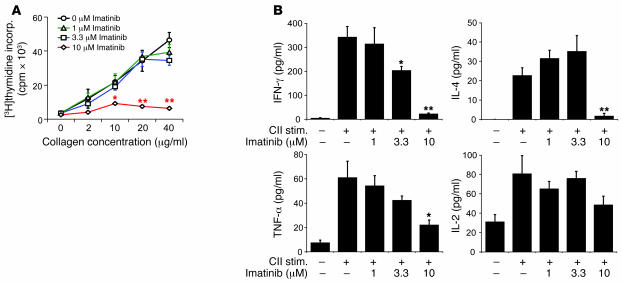
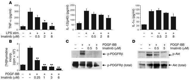
Tyrosine kinases play a central role in the activation of signal transduction pathways and cellular responses that mediate the pathogenesis of rheumatoid arthritis. Imatinib mesylate (imatinib) is a tyrosine kinase inhibitor developed to treat Bcr/Abl-expressing leukemias and subsequently found to treat c-Kit-expressing gastrointestinal stromal tumors. We demonstrate that imatinib potently prevents and treats murine collagen-induced arthritis (CIA). We further show that micromolar concentrations of imatinib abrogate multiple signal transduction pathways implicated in RA pathogenesis, including mast cell c-Kit signaling and TNF-alpha release, macrophage c-Fms activation and cytokine production, and fibroblast PDGFR signaling and proliferation. In our studies, imatinib attenuated PDGFR signaling in fibroblast-like synoviocytes (FLSs) and TNF-alpha production in synovial fluid mononuclear cells (SFMCs) derived from human RA patients. Imatinib-mediated inhibition of a spectrum of signal transduction pathways and the downstream pathogenic cellular responses may provide a powerful approach to treat RA and other inflammatory diseases.


Figures

Figure 1
Figure 1. Imatinib prevents and treats CIA.
(AD) Prevention. DBA/1 mice were administered PBS (n = 15), 33 mg/kg imatinib (n = 15), or 100 mg/kg imatinib (n = 14) orally twice daily starting 1 day prior to induction of CIA. Severity of arthritis was assessed using a visual arthritis scoring system (A) and caliper measurements of paw thickness (B). The incidence of arthritis at the termination of the experiment (day 49) (C) and the mean weights of mice in each group (D) are presented. The data shown in A and B are from a representative of 3 independent experiments, each involving 14–15 mice per experimental arm. (E and F) Treatment. Following the development of clinical arthritis (average visual arthritis score of 4), DBA/1 mice with CIA were randomized and treated with PBS (n = 14), 33 mg/kg imatinib (n = 14), or 100 mg/kg imatinib (n = 14) orally twice daily, and disease was monitored using a visual arthritis scoring system (E) and paw thickness measurements (F). Values from the presented results are the mean ± SEM for this representative experiment. *P < 0.05, **P < 0.01 compared with PBS-treated mice.
Figure 2
Figure 2. Imatinib reduces synovitis, pannus formation, and joint erosions in CIA.
(A) Representative H&E-stained joint tissue sections from DBA/1 mice from a CIA prevention study. (B and C) Histopathological scores of inflammation, pannus formation, and bone and cartilage erosions in DBA/1 mice with CIA in the prevention (B; PBS, n = 8; 33 mg/kg imatinib, n = 8; 100 mg/kg imatinib, n = 8) and treatment (C; PBS, n = 8; 33 mg/kg imatinib, n = 8; 100 mg/kg imatinib, n = 8) studies. Values are mean ± SEM. *P < 0.05, **P < 0.01 compared with PBS-treated group.
Figure 3
Figure 3. Imatinib inhibits mast cell c-Kit activation and proinflammatory cytokine production.
(A) C1.MC/57.1 mast cells were stimulated with 100 ng/ml SCF in the presence of 0–5 μM imatinib, and after 48 hours culture supernatants were collected and analyzed for TNF-α, GM-CSF, and IL-6 by a bead-based cytokine assay. Values are mean ± SEM. *P < 0.05, **P < 0.01 compared with stimulated cells without imatinib. (B and C) C1.MC/57.1 mast cells were serum starved, preincubated with imatinib, and stimulated with 100 ng/ml SCF for 10 minutes in the presence or absence of imatinib, and lysates were generated for IB analysis. IBs were probed with antibodies specific for phospho–c-Kit and total c-Kit (B) and phospho-Akt (Ser473) and total Akt (C). (D) Mast cell lysates generated using the stimulation conditions described in B and C were printed to generate RPP arrays. RPP arrays were probed with a variety of antibodies specific for phosphorylated (activated) protein tyrosine kinases and levels normalized to levels in unstimulated cells. Yellow represents anti-protein tyrosine kinase antibody reactivity, and blue represents lack of reactivity. (E) Mast cells are present in CIA synovium. A representative joint section from a mouse with CIA was stained with toluidine blue. Mast cells present in the densely inflamed CIA synovial tissue are indicated by arrows. B, bone; JS, joint space. Original magnification, ×200.
Figure 4
Figure 4. Inhibition of macrophage c-Fms and downstream MAPK pathways by imatinib.
(A and B) Isolated resident peritoneal macrophages were serum starved, preincubated with imatinib, and stimulated with 100 ng/ml M-CSF for 10 minutes in the presence of imatinib and lysates generated for IB analysis. IBs were probed with antibodies specific for phospho–c-Fms and total Fms (A) or phospho–Akt (Ser473) and total Akt (B). (C) Peritoneal macrophage lysates generated using the stimulation conditions described in A and B were printed on RPP arrays. RPP arrays were probed with a variety of antibodies specific for MAPK pathway and other protein tyrosine kinases, and normalized kinase levels displayed as a heatmap.
Figure 5
Figure 5. Imatinib inhibits B cell proliferation and Ig production in vitro and autoreactive B cell epitope spreading in vivo.
(A) B cells from naive DBA/1 mice were stimulated with 50 μg/ml IgM or 5 μg/ml LPS in the presence of 0–10 μM imatinib. After 48 hours, B cells were pulsed with [3H]thymidine for 18 hours. Data represent mean cpm ± SEM of quadruplicates and are representative of 3 independent experiments. incorp., incorporation. (B) B cells stimulated with LPS (5 μg/ml) were cocultured with 0–10 μM imatinib, and IgM production was measured by ELISA. *P < 0.05, **P < 0.01, ***P < 0.001 compared with stimulated cells without imatinib. (C) Synovial array profiling of serum autoantibodies derived from mice with CIA treated with PBS (n = 7) or 100 mg/kg imatinib (n = 7) (day 49). Synovial microarrays containing candidate autoantigens in RA and CIA were incubated with 1:150 dilutions of mouse sera; autoantibody binding was detected with Cy3-labeled anti-mouse IgG/M secondary antibody; and arrays were scanned to quantify autoantibody binding to each antigen feature. SAM was applied to identify antigen features with statistical differences in autoantibody reactivity in samples derived from PBS-treated mice as compared with imatinib-treated mice (false discovery rate, 0.06). Cluster software was used to order results for the mice and the SAM-identified antigen features, and TreeView software was used to display the resulting clusters of autoantibody reactivity as a heatmap. Red represents positive reactivity, yellow intermediate reactivity, and blue lack of reactivity. Numbers in the key represent digital fluorescence intensity units.
Figure 6
Figure 6. Supratherapeutic imatinib concentrations inhibit T cell responses.
(A) Splenocytes derived from a mouse expressing a transgene encoding a CII-specific TCR were stimulated with 0–40 μg/ml heat-denatured whole CII in the presence of 0–10 μM imatinib. [3H]thymidine incorporation was used to measure proliferation of CII-specific T cells. (B) Bead-based cytokine analysis of culture supernatants from anti-CII TCR transgenic splenocytes stimulated with 20 μg/ml CII from A. Values are mean ± SEM. *P < 0.05, **P < 0.01 compared with stimulated cells without imatinib. stim., stimulation.
Figure 7
Figure 7. Imatinib inhibits SFMC cytokine production and FLS PDGFRβ signaling.
(A) Imatinib inhibits cytokine production by SFMCs derived from a human RA patient. SFMCs were stimulated with 100 ng/ml LPS in the presence of 0–8 μM imatinib, and after 48 hours culture, supernatants were analyzed for TNF-α, IL-12(p40), and IL-1α. Values are mean ± SEM. *P < 0.05 compared with stimulated cells without imatinib. Results are representative of independent experiments performed on SFMCs isolated from 2 RA patients. (B) Modulation of FLS proliferation by imatinib. FLSs from a human RA patient were incubated with 25 ng/ml PDGF-BB in the presence of 0–6 μM imatinib. After 48 hours, FLS cultures were pulsed with [3H]thymidine for 18 hours. Data represent mean cpm ± SEM of quadruplicates and are representative of experiments involving FLS lines derived from 4 RA patients. **P < 0.001 compared with stimulated cells without imatinib. (C and D) Imatinib inhibits PDGFRβ activation in FLS derived from a human RA patient. Cultured FLSs were preincubated with imatinib for 3–4 hours followed by stimulation with 25 ng/ml PDGF-BB for 10 minutes. Lysates were generated and IB analysis performed with antibodies specific for phospho-PDGFRβ and total PDGFRβ (C) and phospho-Akt and total Akt (D). IBs are representative of independent experiments performed on FLS lines derived from 4 RA patients.

References

    1. Firestein G.S. Evolving concepts of rheumatoid arthritis. Nature. 2003;423:356–361. - PubMed
    1. Druker B.J., et al. Efficacy and safety of a specific inhibitor of the BCR-ABL tyrosine kinase in chronic myeloid leukemia. N. Engl. J. Med. 2001;344:1031–1037. - PubMed
    1. Demetri G.D., et al. Efficacy and safety of imatinib mesylate in advanced gastrointestinal stromal tumors. N. Engl. J. Med. 2002;347:472–480. - PubMed
    1. Buchdunger E., Matter A., Druker B.J. Bcr-Abl inhibition as a modality of CML therapeutics. Biochim. Biophys. Acta. 2001;1551:M11–M18. - PubMed
    1. Dewar A.L., et al. Macrophage colony-stimulating factor receptor c-fms is a novel target of imatinib. Blood. 2005;105:3127–3132. - PubMed

Publication types

MeSH terms