

The mechanism of heme transfer from the cytoplasmic heme binding protein PhuS to the delta-regioselective heme oxygenase of Pseudomonas aeruginosa
- PMID: 16981723
- PMCID: PMC2631378
- DOI: 10.1021/bi060980l
The mechanism of heme transfer from the cytoplasmic heme binding protein PhuS to the delta-regioselective heme oxygenase of Pseudomonas aeruginosa
Abstract
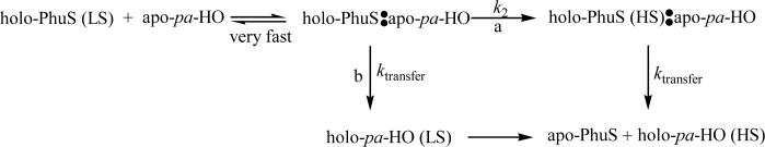
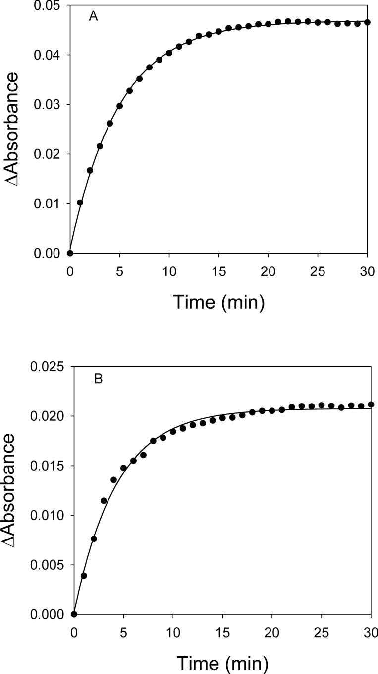
The opportunistic pathogen Pseudomonas aeruginosa has evolved two outer membrane receptor-mediated uptake systems (encoded by the phu and has operons) by which it can utilize the hosts heme and hemeproteins as a source of iron. PhuS is a cytoplasmic heme binding protein encoded within the phu operon and has previously been shown to function in the trafficking of heme to the iron-regulated heme oxygenase (pa-HO). While the heme association rate for PhuS was similar to that of myoglobin, a markedly higher rate of heme dissociation (approximately 10(5) s(-1)) was observed, in keeping with a function in heme-trafficking. Additionally, the transfer of heme from PhuS to pa-HO was shown to be specific and unidirectional when compared to transfer to the non-iron regulated heme oxygenase (BphO), in which heme distribution between the two proteins merely reflects their relative intrinsic affinities for heme. Furthermore, the rate of transfer of heme from holo-PhuS to pa-HO of 0.11 +/- 0.01 s(-1) is 30-fold faster than that to apo-myoglobin, despite the significant higher binding affinity of apo-myoglobin for heme (kH = 1.3 x 10(-8) microM) than that of PhuS (0.2 microM). This data suggests that heme transfer to pa-HO is independent of heme affinity and is consistent with temperature dependence studies which indicate the reaction is driven by a negative entropic contribution, typical of an ordered transition state, and supports the notion that heme transfer from PhuS to pa-HO is mediated via a specific protein-protein interaction. In addition, pH studies, and reactions conducted in the presence of cyanide, suggest the involvement of spin transition during the heme transfer process, whereby the heme undergoes spin change from 6-c LS to 6-c HS either in PhuS or pa-HO. On the basis of the magnitudes of the activation parameters obtained in the presence of cyanide, whereby both complexes are maintained in a 6-c LS state, and the biphasic kinetics of heme transfer from holo-PhuS to pa-HO-wt, supports the notion that the spin-state crossover occur within holo-PhuS prior to the heme transfer step. Alternatively, the lack of the biphasic kinetic with pa-HO-G125V, 6-c LS, and with comparable rate of heme transfer as pa-HO is supportive of a mechanism in which the spin-change could occur within pa-HO. The present data suggests either or both of the two pathways proposed for heme transfer may occur under the present experimental conditions. The dissection of which pathway is physiologically relevant is the focus of ongoing studies.
Figures
References
-
- Wandersman C, Stojiljkovic I. Bacterial heme sources: the role of heme, hemoprotein receptors and hemophores. Curr Opin Microbiol. 2000;3:215–220. - PubMed
-
- Wandersman C, Delepelaire P. Bacterial iron sources: from siderophores to hemophores. Annu Rev Microbiol. 2004;58:611–47. - PubMed
-
- Henderson DP, Payne SM. Cloning and characterization of the Vibrio cholerae genes encoding the utilization of iron from haemin and haemoglobin. Mol Microbiol. 1993;7:461–9. - PubMed
MeSH terms
Substances
Grants and funding
LinkOut - more resources
Full Text Sources
