. 2006 Dec 9;5(12):1439-48.
doi: 10.1016/j.dnarep.2006.07.003. Epub 2006 Sep 18.

NEIL2-initiated, APE-independent repair of oxidized bases in DNA: Evidence for a repair complex in human cells

Affiliations

NEIL2-initiated, APE-independent repair of oxidized bases in DNA: Evidence for a repair complex in human cells

Aditi Das et al. DNA Repair (Amst). .

Abstract

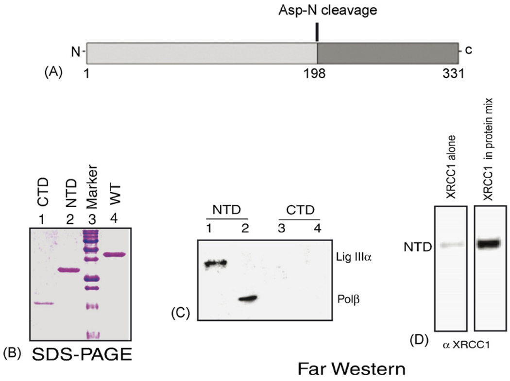
DNA glycosylases/AP lyases initiate repair of oxidized bases in the genomes of all organisms by excising these lesions and then cleaving the DNA strand at the resulting abasic (AP) sites and generate 3' phospho alpha,beta-unsaturated aldehyde (3' PUA) or 3' phosphate (3' P) terminus. In Escherichia coli, the AP-endonucleases (APEs) hydrolyze both 3' blocking groups (3' PUA and 3' P) to generate the 3'-OH termini needed for repair synthesis. In mammalian cells, the previously characterized DNA glycosylases, NTH1 and OGG1, produce 3' PUA, which is removed by the only AP-endonuclease, APE1. However, APE1 is barely active in removing 3' phosphate generated by the recently discovered mammalian DNA glycosylases NEIL1 and NEIL2. We showed earlier that the 3' phosphate generated by NEIL1 is efficiently removed by polynucleotide kinase (PNK) and not APE1. Here we show that the NEIL2-initiated repair of 5-hydroxyuracil (5-OHU) similarly requires PNK. We have also observed stable interaction between NEIL2 and other BER proteins DNA polymerase beta (Pol beta), DNA ligase IIIalpha (Lig IIIalpha) and XRCC1. In spite of their limited sequence homology, NEIL1 and NEIL2 interact with the same domains of Pol beta and Lig IIIalpha. Surprisingly, while the catalytically dispensable C-terminal region of NEIL1 is the common interacting domain, the essential N-terminal segment of NEIL2 is involved in analogous interaction. The BER proteins including NEIL2, PNK, Pol beta, Lig IIIalpha and XRCC1 (but not APE1) could be isolated as a complex from human cells, competent for repair of 5-OHU in plasmid DNA.


Figures

Fig. 1
Fig. 1
Co-immunoprecipitation of NEIL2 and other BER proteins. Extracts of HCT 116 cells transfected with either a FLAG-tagged NEIL2 or control plasmid were immunoprecipitated with anti-FLAG antibody. The blots were then immunoblotted for PNK (A), Lig IIIα (B), XRCC1 (C), Pol β (D) or APE1 (E).
Fig. 2
Fig. 2
Interaction of NEIL2 with BER proteins. (A) Far-Western analysis. Proteins were separated by SDS-PAGE in duplicate gels, one of which was Coomassie stained. The proteins from the other gel were transferred to a nitrocellulose membrane and probed with NEIL2 as described in Section 2. (B) Yeast two-hybrid analysis of NEIL2 and Lig IIIα interaction. Yeast expressing NEIL2 and Lig IIIα fusion proteins (lanes 2 and 4) vs. control expressing only Lig IIIα fusion protein (lanes 1 and 3) were tested on medium with (lanes 1 and 2) and without (lanes 3 and 4) His. Other details are given in Section 2. (C) Right panel, in vitro pulldown of NEIL2 with His-tagged XRCC1 (lane 4) and His-tagged PNK (lane 5), respectively. Left panel, Western analysis using anti-His Ab showing the comparable amounts of His-tagged XRCC1 (lane 1) and PNK (lane 2). (D) Interaction of NEIL2 with full length and fragments of Pol β by Far-Western analysis. Pol β and its truncated fragments (10 µg) were separated by SDS-PAGE and Coomassie-stained (lane 1–6) and tested for interaction with NEIL2 (lanes 7–12). At right, a schematic showing the interacting Pol β domains.
Fig. 3
Fig. 3
(A) Schematic of proteolytic digestion of NEIL2. (B) Coomassie-stained gel of purified C-terminal (CTD, aa residues 199–331), N-terminal domain (NTD, residues 1–198) and WT NEIL2. (C) Far-Western analysis of Lig IIIα and Pol β with NTD (lanes 1 and 2) and CTD (lanes 3 and 4) of NEIL2. (D) Far-Western analysis of NTD of NEIL2 with XRCC1 alone and XRCC1 in protein mix (Pol β, Lig IIIα and PNK).
Fig. 4
Fig. 4
Interaction of NEIL2 with PNK. (A) Extracts of HCT116 cells after co-transfection with expression plasmids of PNK and either vector, FLAG-NEIL2 or FLAG-OGG1 were immunoprecipitated with α-FLAG, and then probed with α-PNK. (A') Reprobing of the blot with α-FLAG. (B) Western analysis of FLAG-immunoprecipitate with α-NEIL2 after cotransfection with non-tagged NEIL2 and FLAG-OGG1, FLAG-PNK or vector alone. (B') Reprobing of the blot with α-FLAG.
Fig. 5
Fig. 5
Repair of 5-OHU in reconstituted system. (A) For α-32PdTMP incorporation into 5-OHU-containing plasmid DNA, 10 pmol substrates were incubated with combinations of 0.5 pmol of NEIL2, 25 fmol of PNK, 25 fmol APE1 and 0.25 pmol of other proteins as described in Section 2. Repaired plasmid was linearized with AhDI and analyzed on 0.8% agarose gel. Lane 1, undamaged DNA and lanes 2–8, 5-OHU-containing plasmid DNA. Complete repair is indicated by the radioactive dTMP incorporation. The agarose gel was dried and the reactions products were analyzed by PhosphorImager. Lane 9, end-labeled 1 kb DNA ladder (NEB). (B) Repair of 5-OHU-containing plasmid DNA with vector (lanes 1 and 2 µl, respectively; 5 and 10 µl, respectively) and FLAG-NEIL2 immunoprecipitate (lanes 3 and 4 µl, respectively; 5 and 10 µl, respectively). Lane 5, end-labeled 1 kb DNA ladder (New England Biolabs).

References

    1. Ames BN. Shigenaga MK. Hagen TM. Oxidants, antioxidants, and the degenerative diseases of aging . Proc. Natl. Acad. Sci. U.S.A. 1993;90:7915–7922. - PMC - PubMed
    1. Breimer LH. Molecular mechanisms of oxygen radical carcinogenesis and mutagenesis: the role of DNA base damage. Mol. Carcinog. 1990;3:188–197. - PubMed
    1. Fraga CG. Shigenaga MK. Park JW. Degan P. Ames BN. Oxidative damage to DNA during aging: 8-hydroxy-2′-deoxyguanosine in rat organ DNA and urine. Proc. Natl. Acad. Sci. U.S.A. 1990;87:4533–4537. - PMC - PubMed
    1. Kreutzer DA. Essigmann JM. Oxidized, deaminated cytosines are a source of C → T transitions in vivo. Proc. Natl. Acad. Sci. U.S.A. 1998;95:3578–3582. - PMC - PubMed
    1. Shibutani S. Takeshita M. Grollman AP. Insertion of specific bases during DNA synthesis past the oxidation-damaged base 8-oxodG. Nature. 1991;349:431–434. - PubMed

Publication types

MeSH terms