Review
. 2006;34(17):4943-59.
doi: 10.1093/nar/gkl620. Epub 2006 Sep 18.

Sequence-specific binding of single-stranded RNA: is there a code for recognition?

Affiliations
Review

Sequence-specific binding of single-stranded RNA: is there a code for recognition?

Sigrid D Auweter et al. Nucleic Acids Res. 2006.

Abstract

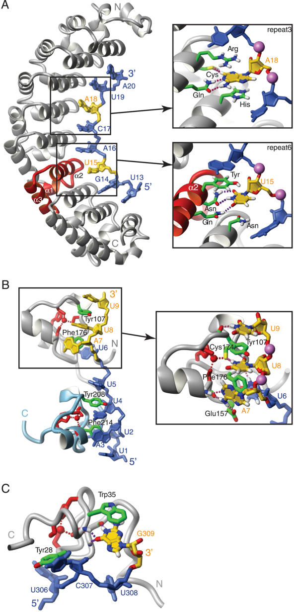
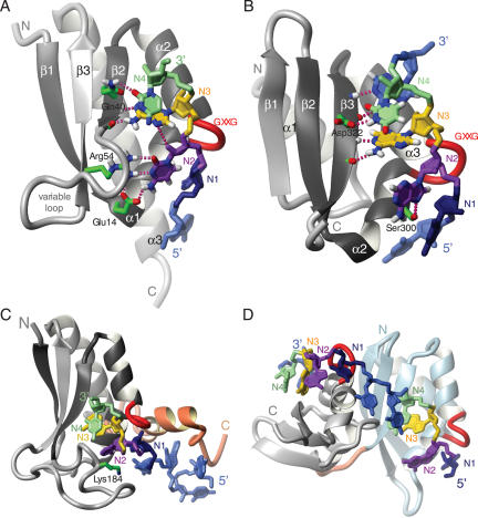
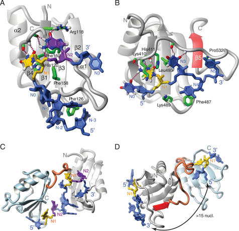
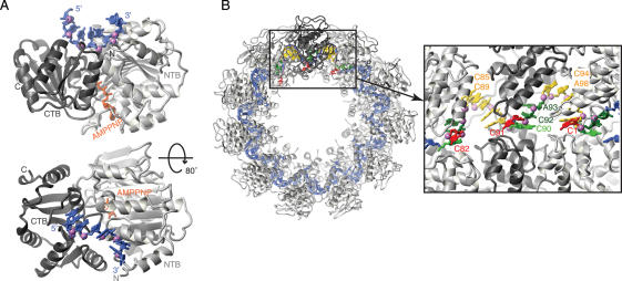
A code predicting the RNA sequence that will be bound by a certain protein based on its amino acid sequence or its structure would provide a useful tool for the design of RNA binders with desired sequence-specificity. Such de novo designed RNA binders could be of extraordinary use in both medical and basic research applications. Furthermore, a code could help to predict the cellular functions of RNA-binding proteins that have not yet been extensively studied. A comparative analysis of Pumilio homology domains, zinc-containing RNA binders, hnRNP K homology domains and RNA recognition motifs is performed in this review. Based on this, a set of binding rules is proposed that hints towards a code for RNA recognition by these domains. Furthermore, we discuss the intermolecular interactions that are important for RNA binding and summarize their importance in providing affinity and specificity.


Figures

Figure 1
Figure 1
Pumilio and zinc-binding domains. (A) Human Pumilio1 in complex with RNA (PDB code: 1M8Y). (B) Complex structure of Tis11d (PDB code: 1RGO). (C) Zinc knuckle of the MMLV nucleocapsid protein in complex with RNA (PDB code: 1U6P). The proteins are shown as grey ribbons; individual protein side-chains are shown in green. Repeat 6 of Pumilio is represented by a red ribbon, the C-terminal zinc finger of Tis11d is represented as a light blue ribbon and the zinc coordinating side-chains in (B and C) are in red. The RNA molecules are in blue and yellow, individual phosphate atoms are shown as purple spheres. Intermolecular hydrogen-bonds are depicted as purple dashed lines. Figures were generated with MOLMOL (88).
Figure 2
Figure 2
KH domains. (A) Type I KH domain of Nova (PDB code: 1EC6). (B) Type II KH domain of NusA (PDB code: 2ATW). (C) KH and QUA2 domains of SF1 (PDB code: 1K1G). (D) Tandem KH domains of NusA (2ATW). The proteins are depicted as grey ribbons, the GXXG loop is shown in red and RNA contacting side-chains are represented by green sticks. The RNA nucleotides N1, N2, N3 and N4 are shown in dark blue, purple, yellow and green, respectively. Other nucleotides are in light blue. Individual intermolecular hydrogen bonds are shown as purple dashed lines. The QUA2 domain of SF1 and the N-terminal KH domain of NusA are shown as red and light blue ribbons. Figures were generated with MOLMOL (88).
Figure 3
Figure 3
RRM domains. (A) The RRM of Fox-1 (PDB code: 2ERR). (B) RRM3 of PTB (PDB code: 2ADC). (C) The tandem RRMs of Sex-lethal (PDB code: 1B7F). (D) RRMs 3 and 4 of PTB (PDB code: 2ADC). The proteins are depicted as grey ribbons, except for the C-terminal RRMs of Sex-lethal and PTB, which are in light blue, and the fifth β-strand of PTB RRM3 and the interdomain linkers, which are in red. Individual side-chains that contact the RNA are represented by green sticks. The RNA nucleotides N1 and N2 are shown in yellow and purple, respectively. Other nucleotides are in blue. Individual hydrogen bonds are shown as purple dashed lines. Figures were generated with MOLMOL (88).
Figure 4
Figure 4
(A) Structures of the DEAD-box protein Vasa (43) and (B) of the rabies virus nucleoprotein (44), two recent non-sequence-specific ssRNA binding proteins in complex with RNA (PDB code: 2DB3 and 2GTT). The protein ribbon is shown as a grey ribbon and the RNA is in dark blue or in color (yellow, green and red) with the phosphate atoms shown as purple spheres. The ATP analogue AMPPNP is shown in orange.
Figure 5
Figure 5
The energies associated with intermolecular stacking interactions. (A) Stacking of U11 and A9 on top of Tyr85 in the MS2 coat protein complex and the effect of Tyr85 mutants on affinity and binding free energy. (B) Contacts between Phe126 and U1, G2 and C3 in the Fox-1 complex and the changes in affinity and binding free energy upon mutating Phe126. (C) Stacking contacts at the U1A RNA binding interface and energetic effects of mutating Phe56. RNA bases are shown in yellow, protein side-chains in green and intermolecular hydrogen bonds as red dashed lines. The table shows dissociation constants (KDs), ratios of KDs and corresponding differences in binding free energy (ΔΔG). Data are taken from (23,50,51). PDB accession codes are 1ZDI, 2ERR and 1URN. Figures were generated with MOLMOL (88).
Figure 6
Figure 6
Arginine and peptide bond stacking. (A) General view and close-up view of the splicing endonuclease in complex with RNA (PDB code: 2GJW) At the splicing endonucleoase active-site, A13 is sandwiched between two arginine side-chains. (B) In the Nova KH domain, N1 stacks on a peptide bond within α1. (C) The N0 nucleotide stacks on a peptide bond that lies at the end of β1 of the RRM of hnRNP A1. The colour scheme is as in Figures 2 and 3. PDB accession codes are 1EC6 (Nova) and 2UP1 (hnRNPA1). Figures were generated with MOLMOL (88).
Figure 7
Figure 7
Surface potential of RNA binding proteins. Blue areas indicate a positive potential, red areas a negative potential. (A) Vts1, a protein that recognizes a structured RNA loop. The RNA binding surface of the protein is a highly positive patch. (B) Fox-1 RRM, which binds ssRNA. Positive and negative potentials surround the RNA and the area where most contacts are made is primarily apolar. Figures were generated with PyMOL () and the surface potential was calculated according to (89). PDB accession codes are 2ESE and 2ERR.
Figure 8
Figure 8
Recognition of AG by hnRNPA1 RRM1. (A) Details of the non-sequence-specific contacts to the RNA. (B) Sequence-specific contacts mediated by the protein main-chain. (C) Sequence-specific contacts mediated by the protein side-chains. The colour scheme is as in Figures 2 and 3. PDB accession code is 2UP1. Figures were generated with MOLMOL (88).

Similar articles

Cited by

References

    1. Klug A. Towards therapeutic applications of engineered zinc finger proteins. FEBS Lett. 2005;579:892–894. - PubMed
    1. Messias A.C., Sattler M. Structural basis of single-stranded RNA recognition. Acc. Chem. Res. 2004;37:279–287. - PubMed
    1. Spassov D.S., Jurecic R. The PUF family of RNA-binding proteins: does evolutionarily conserved structure equal conserved function? IUBMB Life. 2003;55:359–366. - PubMed
    1. de Moor C.H., Meijer H., Lissenden S. Mechanisms of translational control by the 3′ UTR in development and differentiation. Semin. Cell Dev. Biol. 2005;16:49–58. - PubMed
    1. Wang X., McLachlan J., Zamore P.D., Hall T.M. Modular recognition of RNA by a human pumilio-homology domain. Cell. 2002;110:501–512. - PubMed

Publication types