. 2006 Dec;26(23):8892-900.
doi: 10.1128/MCB.01118-06. Epub 2006 Sep 18.

Ubiquitin-binding motifs in REV1 protein are required for its role in the tolerance of DNA damage

Affiliations

Ubiquitin-binding motifs in REV1 protein are required for its role in the tolerance of DNA damage

Caixia Guo et al. Mol Cell Biol. 2006 Dec.

Abstract

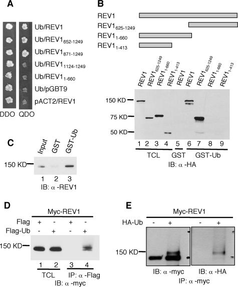
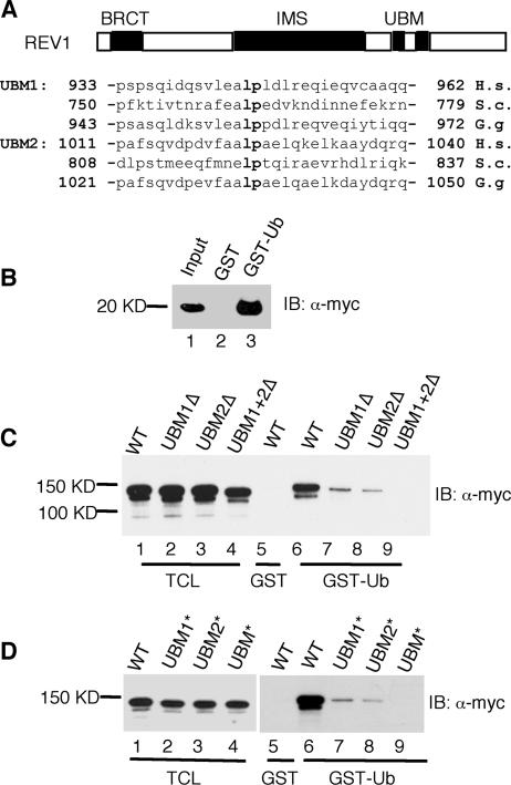
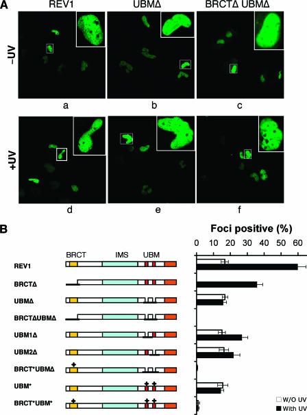
REV1 protein is a eukaryotic member of the Y family of DNA polymerases involved in the tolerance of DNA damage by replicative bypass. The precise role(s) of REV1 in this process is not known. Here we show, by using the yeast two-hybrid assay and the glutathione S-transferase pull-down assay, that mouse REV1 can physically interact with ubiquitin. The association of REV1 with ubiquitin requires the ubiquitin-binding motifs (UBMs) located at the C terminus of REV1. The UBMs also mediate the enhanced association between monoubiquitylated PCNA and REV1. In cells exposed to UV radiation, the association of REV1 with replication foci is dependent on functional UBMs. The UBMs of REV1 are shown to contribute to DNA damage tolerance and damage-induced mutagenesis in vivo.


Figures

FIG. 1.
FIG. 1.
Interaction between REV1 and ubiquitin. (A) Full-length and deletion mutants of REV1 cloned in pGBT9 vector were tested for the ability to interact with Ub-pACT2 in the yeast two-hybrid system. Yeast strain AH109 was cotransformed with plasmid combinations as indicated and plated on auxotroph-selective medium (QDO). The presence of “bait” and “prey” plasmids in cotransformed cells was controlled by growth on double-drop-out medium (DDO). (B) Cos7 cell lysates expressing full-length and truncated REV1 proteins tagged with HA epitopes at their N termini were incubated with GST or GST-ubiquitin fusion proteins as indicated. Bound proteins were detected by immunoblot analysis with anti-HA (α-HA) antibody. The TCL lanes (lane 1 to 4) contain 1/50 of the lysates used in the experiments. IB, immunoblot. (C) GST-ubiquitin pull-down assay of purified REV1 protein. Lane 1, input containing 1/10 of the REV1 used in the experiments; lane 2, GST plus REV1; lane 3, GST-Ub plus REV1. (D) α-Flag M2 agarose affinity gel was incubated with the Cos7 cell lysates expressing Myc-REV1 and Flag-Ub or Flag (control), as indicated. Lanes 1 and 2, input containing 1/50 of the lysates used for immunoprecipitation; lanes 3 and 4, immunoprecipitation of lysates with α-Flag. (E) HEK293T cells were transfected with (+) or without (−) HA-tagged ubiquitin (HA-Ub) together with Myc-tagged REV1 constructs. The lysates were immunoprecipitated with α-Myc antibody and subjected to immunoblotting with α-Myc and α-HA as indicated.
FIG. 2.
FIG. 2.
The UBMs in REV1 are required for association between REV1 protein and ubiquitin. (A) Schematic representation of REV1 protein domains and the amino acid sequences of the UBMs. (B) Cos7 cell lysates expressing Myc-REV1-UBM1 + UBM2 were pulled down with GST or GST-ubiquitin fusion proteins as indicated. Bound proteins were detected by immunoblot analysis with anti-Myc (α-Myc) antibody. The input contains 1/50 of the lysates used in the experiment. (C and D) Cos7 cell lysates expressing full-length REV1 and REV1 with the UBMs deleted (C) or REV1 with the UBMs mutated (D). Myc-tagged REV1 proteins were pulled down with GST or GST-ubiquitin fusion proteins as indicated. Bound proteins were detected by immunoblot analysis with α-Myc. The TCL lanes (lanes 1 to 4) contain 1/50 of the lysates used in the experiments.
FIG. 3.
FIG. 3.
The UBMs in REV1 protein mediate increased association between REV1 and monoubiquitylated PCNA. (A) The UBMs in REV1 mediate interaction with monoubiquitylated PCNA (mUb-PCNA). MRC5 cells were UV irradiated with 25 J/m2; 6 to 8 h later, the Triton-insoluble fractions were harvested and incubated with GST-REV1923-1150 (REV1) or its UBM mutant (REV1-U*) as indicated. Retained proteins were detected by immunoblotting with anti-PCNA (α-PCNA). A small amount of nonubiquitylated PCNA and another band on top of the monoubiquitylated form (according to size, it is likely diubiquitylated PCNA) precipitated with GST-REV1923-1150, presumably due to heterotrimerization of PCNA containing a mixture of ubiquitylated and nonubiquitylated forms. (B) GST-REV1923-1150 (REV1) or its UBM mutant (REV1-U*) pulled down purified PCNA-Ub protein as indicated. Retained proteins were detected by immunoblotting with α-PCNA. The input lanes contain 1/10 of the purified proteins used in the experiments. (C and D) Lysates of Cos7 cells expressing wild-type (W) and UBM* (U*) REV1 protein (C) or wild-type (W) and BRCT* UBM* (M) REV1 protein (D) tagged with GFP epitope at their N termini were incubated with GST fusion proteins as indicated. Bound proteins were detected by immunoblotting with α-GFP. The TCL lanes contain 1/75 of the lysates used in the experiments. (E) HEK293T cells were transfected with wild-type (W) or BRCT* UBM* (M) GFP-REV1, and 40 h later they were UV irradiated (25 J/m2). They were then incubated for 7 h prior to Triton extraction and cross-linking. Triton-insoluble proteins were solubilized and immunoprecipitated with α-PCNA, and the retained proteins were analyzed by Western blotting with α-GFP (top) or α-PCNA (bottom). Input lanes included Triton-soluble fractions (SF) and insoluble fractions (IF). Data are representative of three independent experiments.
FIG. 4.
FIG. 4.
UBMs are required for optimal association of REV1 with replication factories in cells exposed to UV radiation. (A) MRC5 cells were transfected with plasmids encoding full-length or deletion mutants of eGFP-REV1 as indicated. Twenty hours after transfection, cells were UV irradiated (10 J/m2) (bottom panel) and incubated for 8 to 16 h before fixation with paraformaldehyde. The distribution of REV1 or deletion mutants (as indicated) was observed directly by autofluorescence of eGFP. The images show unirradiated (top) or UV-irradiated (bottom) transfected cells. (B) REV1 focus formation after UV irradiation. MRC5 cells were transfected with a panel of eGFP-REV1 mutants and incubated for 20 h. Cells were irradiated with 10 J/m2 UVC radiation and further incubated for 8 h. The proportion of eGFP-REV1 expressing cells in which the protein was localized in nuclear foci was determined. All experiments were carried out in triplicate. Error bars indicate the standard error. Note that the result for BRCTΔ cells was extracted from reference .
FIG. 5.
FIG. 5.
The UBMs and the BRCT domain of REV1 contribute independently to the tolerance to UV and CDDP in DT40. Δrev1 clones containing each mutant REV1 cDNA were established. The UV and CDDP sensitivities of the wild type, Δrev1, and each mutant clone were measured by clonogenic colony survival assays. The data shown are representative results from three independent transformants, which exhibited almost the same sensitivities. Data points show the average and standard deviation of triplicate experiments for each clone.
FIG. 6.
FIG. 6.
UBM2 of REV1 contributes to efficient PCNA-dependent lesion bypass in S. cerevisiae. (A) The function of yeast REV1 depends on modification of PCNA (encoded by POL30) at K164. Epistasis analysis was carried out by comparing the sensitivities toward MMS and NQO of a rev1 deletion and of the pol30-K164R mutant with those of the double mutant. Plates were incubated 2 or 3 days to assess growth at the indicated toxin concentrations. (B) UBM2 of yeast contributes to lesion bypass. Damage sensitivities were analyzed as above with a series of strains constructed by introducing appropriate alleles of yeast REV1 on integrative plasmids into a rev1 deletion strain. rev1, empty expression vector; REV1, wild-type REV1 allele; UBM1*, L763A P764A; UBM2*, L821A P822A; UBM*, L763A P764A L821A P822A; UBM* BRCT*, L763A P764A L821A P822A G193R. (C) Quantification of MMS sensitivities by determination of survival after treatment with 0.1% MMS for various times. (D) Effects of REV1 mutations on UV sensitivity. Symbols are used as in panel C. (E) Effects of REV1 mutations on MMS-induced mutagenesis. The number of canavanine-resistant (Canr) mutants per 106 surviving cells in response to various doses of MMS was determined by plating aliquots of treated cultures on YPD to determine viable cell numbers and on canavanine-containing plates for measuring the number of mutants. Symbols are used as in panel C. Standard deviations were calculated from triplicate measurements.

References

    1. Baynton, K., A. Bresson-Roy, and R. P. Fuchs. 1999. Distinct roles for Rev1p and Rev7p during translesion synthesis in Saccharomyces cerevisiae. Mol. Microbiol. 34:124-133. - PubMed
    1. Bebenek, K., and T. A. Kunkel. 2004. Functions of DNA polymerases. Adv. Protein Chem. 69:137-165. - PubMed
    1. Bienko, M., C. M. Green, N. Crosetto, F. Rudolf, G. Zapart, B. Coull, P. Kannouche, G. Wider, M. Peter, A. R. Lehmann, K. Hofmann, and I. Dikic. 2005. Ubiquitin-binding domains in Y-family polymerases regulate translesion synthesis. Science 310:1821-1824. - PubMed
    1. Buerstedde, J. M., and S. Takeda. 1991. Increased ratio of targeted to random integration after transfection of chicken B cell lines. Cell 67:179-188. - PubMed
    1. Friedberg, E. C., A. R. Lehmann, and R. P. Fuchs. 2005. Trading places: how do DNA polymerases switch during translesion DNA synthesis? Mol. Cell 18:499-505. - PubMed

Publication types

MeSH terms