. 2006 Nov;26(22):8527-38.
doi: 10.1128/MCB.01035-06. Epub 2006 Sep 18.

Two domains of the erythropoietin receptor are sufficient for Jak2 binding/activation and function

Affiliations

Two domains of the erythropoietin receptor are sufficient for Jak2 binding/activation and function

Stéphane Pelletier et al. Mol Cell Biol. 2006 Nov.

Abstract

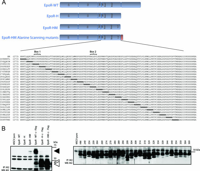
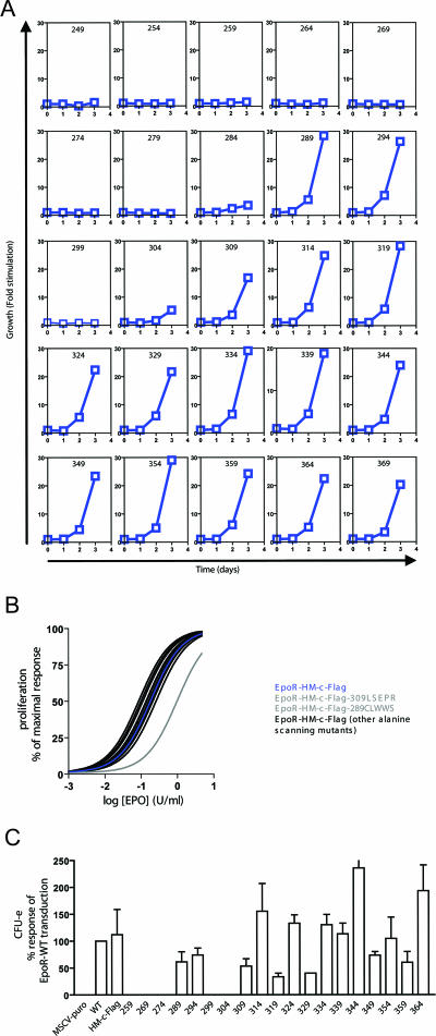
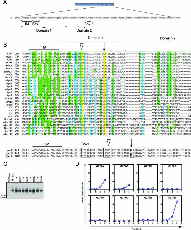
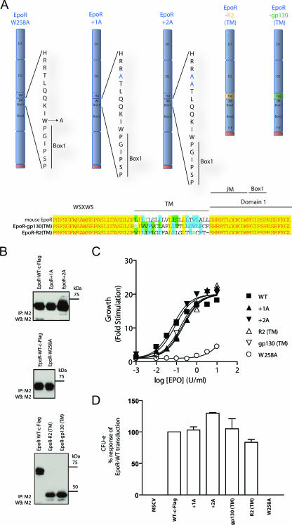
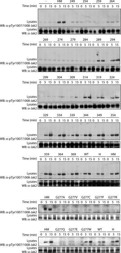
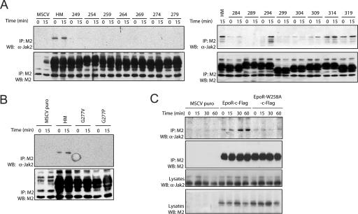
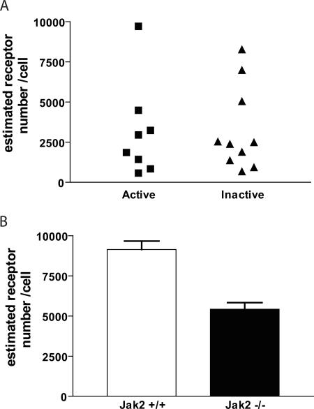
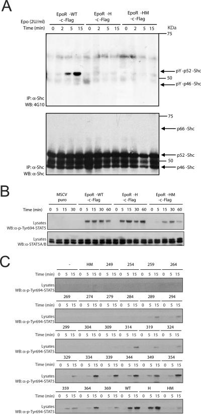
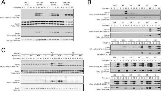
Biochemical and genetic studies have shown that Jak2 is an essential component of EpoR signal transduction which is required for normal erythropoiesis. However, whether Jak2 is the sole direct mediator of EpoR signal transduction remains controversial. To address this issue, we have used an extensive and systematic mutational analysis across the EpoR cytoplasmic tail and transmembrane domain with the goal of determining whether mutants that negatively affected EpoR biological activity but retained Jak2 activation could be identified. Analysis of over 40 mutant receptors established that two large domains in the membrane-proximal region, which include the previously defined Box1 and Box2 domains as well as a highly conserved glycine among cytokine receptors, are required for Jak2 binding and activation and to sustain biological activity of the receptor. Importantly, none of the mutants that lost the ability to activate Jak2 retained the ability to bind Jak2, thus questioning the validity of models of receptor reorientation for Jak2 activation. Also, no correlation was made between cell surface expression of the receptor and its ability to bind Jak2, thus questioning the role of Jak2 in trafficking the receptor to the plasma membrane. Collectively, the results suggest that Jak2 is the sole direct signaling molecule downstream of EpoR required for biological activity.


Figures

FIG. 1.
FIG. 1.
Outline of the WT and the H, HM, and alanine scanning mutant receptors. (A) Schematic representation of the WT and the H and HM mutant receptors and sequence alignment of the various alanine scanning mutants. D1, extracellular domain 1, which is required for Epo binding; D2, extracellular domain 2, which is required for Epo binding; JM, juxtamembrane domain; Box1, highly conserved amino acid sequence in cytokine receptor family; Box2, highly conserved amino acid sequence in cytokine receptors; Y-F, Y343F. The alanine scanning mutants are named based on the number of the first amino acid changed by an alanine. The number in parentheses is the amino acid position in the alignment for the immature form of the polypeptide. (B) Whole-cell extracts, prepared from exponentially growing DA3 cell lines expressing or not expressing (pMSCVpuro) the various EpoR constructs, were subjected to immunoprecipitation (IP) using the anti-Flag antibody (M2) and analyzed by Western blotting (WB) using the M2 antibody. EpoR-WT, DA3 cells expressing WT EpoR; EpoR-WT-c-Flag, DA3 cell line expressing WT-c-Flag EpoR; 249, DA3 cell line expressing the EpoR-HM mutant 249; etc.
FIG. 2.
FIG. 2.
Two functional domains are required for the biological activity of EpoR. (A) Exponentially growing DA3 cell lines were starved for 12 to 16 h and then treated with Epo (5 U/ml). Cell numbers were counted every 24 h over a period of 3 days. The results are expressed as increase in cell number (stimulation [n-fold]) compared to the number of cells plated on day 0 (for Epo stimulation) and represent the means of triplicate results for each time point and each cell line. (B) Exponentially growing DA3 cell lines that responded to Epo (as presented in panel A) were starved for 12 to 16 h and then stimulated with increasing concentrations of Epo (0.001 to 5 U/ml) for 3 days. Cell numbers were counted after 3 days. The results are expressed in percentages of maximal response and represent the means of triplicate results for each concentration. (C) EpoR-deficient fetal liver cells were infected with empty or EpoR-expressing retroviruses and subjected to in vitro colony assays with Epo (5 U/ml). Benzidine-positive CFU-e were scored at days 2 to 3. Results are expressed as the percentages of colonies observed compared to WT EpoR transduction and represent the means ± standard errors of the means from three independent experiments. Descriptions of designations are given in the legend for Fig. 1.
FIG. 3.
FIG. 3.
Domain 1 contains a highly conserved glycine which is essential for biological activity. (A) Schematic representation of domains 1 and 2, required for biological activity of the receptor. Abbreviations are defined in the legend for Fig. 1A. (B) Sequence alignment of various cytokine receptors and species variants, illustrating the high degree of conservation of glycine 277 (black arrow) and proline 262 (open arrowhead). Abbreviations: GHR, growth hormone receptor; PRLR, prolactin receptor; TSLPR, thymic stromal lymphopoietin receptor. Prefixes: h, Homo sapiens; m, Mus musculus; x, Xenopus laevis; ss, Sus scrofa; cerf, Cervus elaphus; dr, Danio rerio; oa, Ovis aries; r, Rattus norvegicus; t, Tetraodon nigroviridis; c, Canis familiaris; b, Bos taurus. (C) Whole-cell extracts prepared from exponentially growing DA3 cell lines expressing or not expressing (pMSCVpuro) the various EpoR constructs were subjected to immunoprecipitation (IP) using the anti-Flag antibody (M2) and analyzed by Western blotting (WB) using the M2 antibody. (D) See the legend for Fig. 2A for details.
FIG. 4.
FIG. 4.
Outline and biological activities of the W258A, +1A, and +2A EpoR mutants and the EpoR-R2(TM) and EpoR-gp130(TM) transmembrane swap mutants. (A) Schematic representation of W258A, +1A, +2A, EpoR-R2(TM), and EpoR-gp130(TM) mutants and sequence alignment of EpoR compared to the transmembrane swap mutants. (B) Whole-cell extracts, prepared from exponentially growing DA3 cell lines expressing or not expressing (pMSCVpuro) the various EpoR constructs, were subjected to immunoprecipitation (IP) using the anti-Flag antibody (M2) and analyzed by Western blotting (WB) using the M2 antibody. (C) Exponentially growing DA3 cells were starved for 12 to 16 h and then stimulated with increasing concentrations of Epo (0.001 to 10 U/ml) for 3 days. Cell numbers were counted after 3 days, and the results are expressed as the increase (n-fold) in cell number. (D) EpoR-deficient fetal liver cells were infected with empty or EpoR-expressing retroviruses and subjected to in vitro colony assays with Epo (5 U/ml). Benzidine-positive CFU-e were scored at days 2 and 3. Results are expressed as the percentages of colonies observed compared to WT EpoR transduction and represent the means ± standard errors of the means from three independent experiments. Designations and abbreviations are defined in the legend for Fig. 1.
FIG. 5.
FIG. 5.
Correlation between Jak2 activation and biological activity of the receptor. DA3 cell lines expressing the various alanine scanning and G277 mutants were starved for 12 to 16 h and then stimulated with Epo (1 U/ml) for the indicated times. Whole-cell extracts were analyzed by immunoblotting using an anti-phospho-Y1007/1008 Jak2 antibody. The same extracts were also analyzed by immunoblotting using an anti-Jak2 antibody. WB, Western blotting.
FIG. 6.
FIG. 6.
Correlation between Jak2 binding and biological activity of the receptor. IL-3-deprived DA3 cell lines expressing or not expressing (pMSCVpuro or MSCV) the EpoR-HM mutant (HM) and various biologically inactive and some active alanine scanning mutants (A), G277 mutants (B), and the W258 mutant (C) were left untreated or treated with Epo (1 U/ml) for 15, 30, or 60 min before solubilization. Cell lysates were subjected to immunoprecipitation (IP) using the anti-Flag antibody (M2). EpoR and associated Jak2 were analyzed by Western blotting (WB) using the M2 and anti-Jak2 antibodies, respectively.
FIG. 7.
FIG. 7.
Jak2 association with EpoR does not affect EpoR cell surface expression. (A) Cell surface expression of biologically active (WT, H, HM, +1A, +2A, 289, 294, and 309 mutants) or inactive (249, 254, 264, 269, 274, 279, 284, 299, 304, and W258A mutants) mutant receptors in DA3 cells. (B) Cell surface expression of WT EpoR in WT or Jak2-deficient MEFs. Receptor density at the cell surface was determined as described in Materials and Methods, and results are expressed as means ± standard errors of the means.
FIG. 8.
FIG. 8.
Proximal downstream signaling from EpoR mutants. (A) IL-3-deprived DA3 cell lines expressing WT EpoR and the EpoR-H and EpoR-HM mutants were left untreated or treated with Epo (2 U/ml) for the indicated times before solubilization. Cell lysates were subjected to immunoprecipitation (IP) with an anti-Shc antibody, and tyrosine phosphorylation was analyzed by Western blotting (WB) using antiphosphotyrosine antibody 4G10. Immunoprecipitated Shc proteins were also analyzed by Western blotting using an anti-Shc antibody. (B and C) IL-3-deprived DA3 cell lines expressing WT EpoR and the EpoR-H and EpoR-HM mutants (B) and alanine scanning mutants (C) were left untreated or treated with Epo (1 U/ml) for the indicated times. The activation of Stat5 was monitored by immunoblotting with phosphospecific antibodies against the activating tyrosine phosphorylation site (Tyr694). Descriptions of designations are given in the legend for Fig. 1.
FIG. 9.
FIG. 9.
Distal downstream signaling from EpoR. IL-3-deprived DA3 cell lines expressing WT EpoR and the EpoR-H and EpoR-HM mutants (A) and alanine scanning mutants (B) were left untreated or treated with Epo (1 U/ml) for the indicated times. The activating phosphorylation of Erk1/Erk2 and Akt was monitored by immunoblotting of lysate proteins with phosphospecific antibodies as indicated. (C) Exponentially growing Jak2−/− MEFs stably expressing or not expressing Jak2-WT-c-HA or the kinase-inactive Jak2-KD-c-HA mutant (KD, kinase dead) and with or without EpoR-WT-c-Flag and HM mutants were starved for 12 to 16 h and then stimulated with Epo for the indicated times. The activating phosphorylation of Erk1/Erk2 and Akt was monitored by immunoblotting of lysate proteins with phosphospecific antibodies as indicated. WB, Western blotting. Descriptions of designations are given in the legend for Fig. 1.

Similar articles

Cited by

References

    1. Bonifacino, J. S. 2002. Quality control of receptor-kinase signaling complexes. Dev. Cell 2:1-2. - PubMed
    1. Constantinescu, S. N., L. J. Huang, H. Nam, and H. F. Lodish. 2001. The erythropoietin receptor cytosolic juxtamembrane domain contains an essential, precisely oriented, hydrophobic motif. Mol. Cell 7:377-385. - PubMed
    1. He, K., X. Wang, J. Jiang, R. Guan, K. E. Bernstein, P. P. Sayeski, and S. J. Frank. 2003. Janus kinase 2 determinants for growth hormone receptor association, surface assembly, and signaling. Mol. Endocrinol. 17:2211-2227. - PubMed
    1. Hofmann, S. R., A. Q. Lam, S. Frank, Y.-J. Zhou, H. L. Ramos, Y. Kanno, D. Agnello, R. J. Youle, and J. J. O'Shea. 2004. Jak3-independent trafficking of the common γ chain receptor subunit: chaperone function of Jaks revisited. Mol. Cell. Biol. 24:5039-5049. - PMC - PubMed
    1. Huang, L. J., S. N. Constantinescu, and H. F. Lodish. 2001. The N-terminal domain of Janus kinase 2 is required for Golgi processing and cell surface expression of erythropoietin receptor. Mol. Cell 8:1327-1338. - PubMed

Publication types

LinkOut - more resources