. 2006 Nov;26(22):8448-60.
doi: 10.1128/MCB.01248-06. Epub 2006 Sep 18.

Specific isoforms of translation initiation factor 4GI show differences in translational activity

Affiliations

Specific isoforms of translation initiation factor 4GI show differences in translational activity

Mark J Coldwell et al. Mol Cell Biol. 2006 Nov.

Abstract

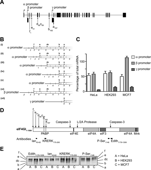
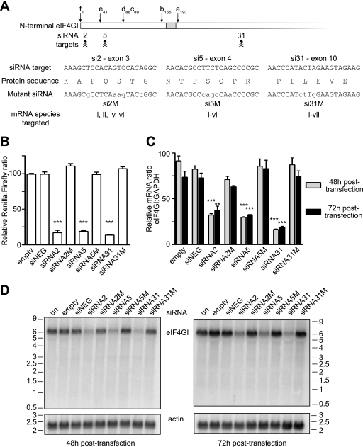
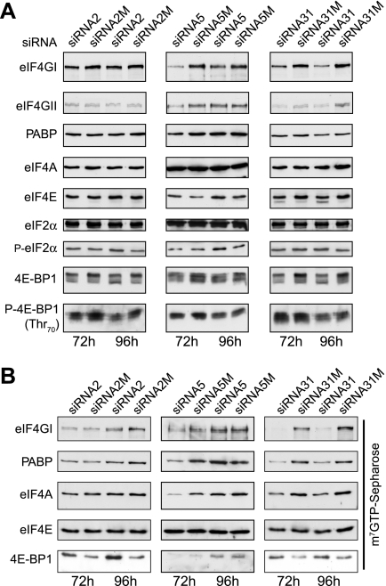
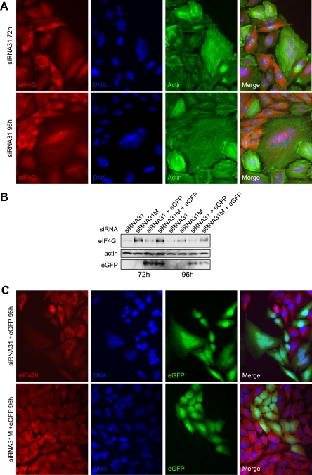
The eukaryotic initiation factor (eIF) 4GI gene locus (eIF4GI) contains three identified promoters, generating alternately spliced mRNAs, yielding a total of five eIF4GI protein isoforms. Although eIF4GI plays a critical role in mRNA recruitment to the ribosomes, little is known about the functions of the different isoforms, their partner binding capacities, or the role of the homolog, eIF4GII, in translation initiation. To directly address this, we have used short interfering RNAs (siRNAs) expressed from DNA vectors to silence the expression of eIF4GI in HeLa cells. Here we show that reduced levels of specific mRNA and eIF4GI isoforms in HeLa cells promoted aberrant morphology and a partial inhibition of translation. The latter reflected dephosphorylation of 4E-BP1 and decreased eIF4F complex levels, with no change in eIF2alpha phosphorylation. Expression of siRNA-resistant Myc-tagged eIF4GI isoforms has allowed us to show that the different isoforms exhibit significant differences in their ability to restore translation rates. Here we quantify the efficiency of eIF4GI promoter usage in mammalian cells and demonstrate that even though the longest isoform of eIF4GI (eIF4GIf) was relatively poorly expressed when reintroduced, it was more efficient at promoting the translation of cellular mRNAs than the more highly expressed shorter isoforms used in previous functional studies.


Figures

FIG. 1.
FIG. 1.
The three eIF4GI promoters are used with different efficiencies in HeLa cells. A. Diagrammatic representation of the eIF4GI gene locus, showing the locations of the three promoters, designated α, β, and γ. Coding exons are shown in gray, and noncoding exons are shown in black. B. Diagrammatic representation of the eIF4GI mRNAs showing alternative splice variants generated from the different promoters (adapted from reference 10). C. Total RNA from the cell types shown was reverse transcribed and then amplified with primers specific to eIF4GI mRNAs. To quantify mRNAs arising from the α promoter, a primer pair that amplified only sequences containing exon 1 was created. The amplicon to quantify the mRNAs arising from the β promoter utilized a forward primer with complementarity to the β exon and a reverse primer specific for exon 3, while the γ promoter activity was quantified with a primer pair that amplified mRNAs containing exons γ and 9. Finally, a forward primer specific for exon 28 and a reverse primer complementary to exon 29 were used to quantify the total amount of eIF4GI mRNA in cells. Values obtained by quantitative RT-PCR were normalized to those obtained from an amplicon which detected all eIF4GI mRNA. Data were collected from three independent experiments, with assays each performed in triplicate, and error bars represent the standard errors of the means. D. Schematic representation of the eIF4GI protein. This figure shows the sites of alternative translation initiation, binding sites of other components of the translation initiation machinery (7, 10, 36), and epitopes used to generate antisera used in these studies. Sites of cleavage by caspase-3 and the picornaviral (L/2A) proteases are indicated. E. Equal amounts of extract from the cell types indicated were subjected to SDS-PAGE using 4% acrylamide precast gels and proteins transferred to PVDF. The membrane was then probed with the antibodies shown to visualize the eIF4GI isoforms (indicated on the left and right of the figure).
FIG. 2.
FIG. 2.
Plasmid-derived siRNA can be used to knock down eIF4GI mRNA in HeLa cells. A. Three siRNA target sites were identified in the N terminus of the eIF4GI sequence, and corresponding oligonucleotides (si2, si5, and si31) were inserted into the pSilencer 3.0 H1 plasmid (Ambion). Mutated, control versions of each siRNA (si2M, si5M, and si31M) were also created that contained at least three mismatches to the center of the target site. B. siRNAs directed against eIF4GI specifically inhibit the expression of a Renilla luciferase mRNA fused to the eIF4GI N-terminal RNA sequence eIF4GI in the psiCHECK-2 vector. Dual-luciferase assays were performed on HeLa cell extracts prepared after 48 h of transfection, and the Renilla luciferase activity was normalized to firefly luciferase expressed from the same plasmid. Each variable was compared with data from cells transfected with the plasmid expressing a negative control siRNA (siNEG); ***, P < 0.001; n = 3, with error bars representing the standard errors of the means. C. HeLa cells were transfected with siRNA-expressing plasmids for the times shown, and total RNA was extracted and reverse transcribed, as described. The presence of eIF4GI or GAPDH mRNA was measured by quantitative RT-PCR. Each variable was compared as described for panel B; ***, P < 0.001; **, P < 0.01; n = 3, with error bars representing the standard errors of the means. D. Total RNA from HeLa cells, transfected with the indicated plasmids for the times shown, was subjected to denaturing agarose gel electrophoresis in the presence of formaldehyde. RNA was transferred to nitrocellulose and then probed with a radiolabeled, single-stranded DNA specific for the C terminus of eIF4GI. The blot was subsequently reprobed with a radiolabeled probe specific for β-actin.
FIG. 3.
FIG. 3.
Silencing of eIF4GI mRNA with alternative siRNAs has differing effects on eIF4GI protein expression and translation rates. A. Extracts prepared from HeLa cells after transfection for 72 h (left panel) or 96 h (right panel) with the siRNA plasmids indicated were subjected to SDS-PAGE, and proteins were transferred to PVDF, with eIF4GI and actin visualized by immunoblotting. un refers to untransfected cells, and empty refers to cells transfected with the vector alone. B. Prior to the harvest after 96 h of transfection, cells were incubated with [35S]methionine for 1 h, extracts prepared, and the incorporation of radioactive methionine into protein determined as described in the text (expressed as cpm/μg protein). *, P < 0.05; **, P < 0.01; ns, not significant, P = >0.05; n = 3, with error bars representing the standard errors of the means. C. Equal amounts of extract from HeLa cells transfected for 72 h or 96 h with the indicated siRNA plasmids were resolved by SDS-PAGE using 10% acrylamide gels (upper and lower panels) of 4% acrylamide precast gels (middle panel). eIF4GI was visualized by immunoblotting, as described, and the migration of the different isoforms is indicated on the right. D. To determine promoter activity in cells transfected with siRNA2 and siRNA5, total RNA was obtained from cells transfected for the times indicated and subjected to reverse transcription. Quantitative RT-PCR was performed to amplify sequences derived from each promoter for total eIF4GI mRNA and for GAPDH mRNA. The values obtained are expressed relative to the amounts of each amplicon present in untransfected cells (set at 100). Data were collected from three independent experiments, with assays each performed in triplicate, and error bars represent the standard errors of the means.
FIG. 4.
FIG. 4.
Silencing of eIF4GI also results in reduced levels of eIF4E and the dephosphorylation of 4E-BP1. A. Equal amounts of cell extract from HeLa cells transfected with the indicated siRNA plasmids for 72 h or 96 h were subjected to SDS-PAGE and the indicated proteins visualized by immunoblotting. B. Aliquots of extracts containing equal amounts of protein were subjected to m7GTP-Sepharose affinity chromatography to recover eIF4E and associated factors. Proteins were resolved by SDS-PAGE and visualized by immunoblotting using the antisera indicated. These data are from a single experiment but are representative of those obtained in at least three separate experiments.
FIG. 5.
FIG. 5.
HeLa cells with reduced expression of eIF4GI often show a multinucleated morphology. A. HeLa cells were grown on glass coverslips and after transfection for 72 h (top panels) or 96 h (bottom panels) with siRNA31 were processed for immunofluorescence microscopy, as described in the text. eIF4GI was detected with the C-terminus-specific antibody, followed by goat antirabbit immunoglobulin G conjugated to tetramethyl rhodamine isocyanate; actin was detected with phalloidin conjugated to FITC, while nuclei were stained with 4′,6′-diamidino-2-phenylindole hydrochloride. These data are from a single experiment but are representative of those obtained in at least five separate experiments. B. HeLa cells were transfected with siRNA31 expression vectors (lanes 1, 2, 5, and 6) or siRNA31-eGFP vectors (lane 3, 4, 7, and 8) for the times indicated. Extracts were prepared, and eIF4GI, actin, and eGFP expression was monitored by SDS-PAGE and immunoblotting, as indicated. C. HeLa cells were grown on glass coverslips and after transfection for 96 h with siRNA31-eGFP (upper panels) or siRNA31M-eGFP (lower panels) were processed for immunofluorescence microscopy, as described for panel A. These data are from a single experiment but are representative of those obtained in at least three separate experiments.
FIG. 6.
FIG. 6.
Low levels of eIF4GIf are more efficient than high levels of eIF4GIa at partial restoration of translation rates following reduced expression of endogenous eIF4GI. A (upper). HeLa cells were cotransfected for 96 h with siRNA vectors and plasmids expressing different Myc-tagged isoforms of eIF4GI or the apoptotic cleavage fragment of eIF4G (M-FAG), as indicated. Cell extracts were prepared and aliquots containing equal amounts of total protein resolved by SDS-PAGE using 10% acrylamide gels or 4% acrylamide precast gels, as indicated. eIF4GI was visualized by immunoblotting using either anti-Myc antibody or anti-eIF4GI antiserum, and other proteins were visualized using the antisera indicated. (Lower) Aliquots of extracts containing equal amounts of protein were subjected to m7GTP-Sepharose affinity chromatography to recover eIF4E and associated factors. Proteins were resolved by SDS-PAGE and visualized by immunoblotting using the antisera indicated. B (upper). HeLa cells were cotransfected for 96 h with siRNA vectors and plasmids expressing Myc-eIF4GII, as indicated. Extracts were prepared and proteins visualized as for panel A. (Lower) Aliquots of extracts containing equal amounts of protein were subjected to m7GTP-Sepharose affinity chromatography to recover eIF4E and associated factors, as described above. C. Prior to the harvest after 96 h of transfection, cells were incubated with [35S]methionine for 1 h, extracts prepared, and the incorporation of radioactive methionine into protein determined as described in the text (expressed as cpm/μg total protein). Unpaired two-way t tests were performed separately, comparing eIF4GIf with si31 (formula imageformula imageformula image
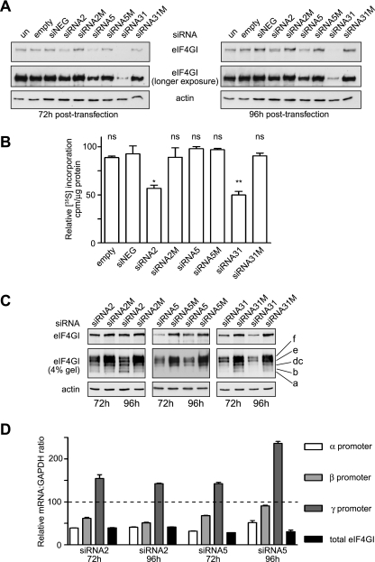
, P < 0.001; n = 3) and comparing eIF4GIf with other isoforms of eIF4GI (formula imageformula image, P < 0.01; ns, P = >0.05; n = 3), with error bars representing standard errors of the means.

Similar articles

Cited by

References

    1. Abbott, C. M., and C. G. Proud. 2004. Translation factors: in sickness and in health. Trends Biochem. Sci. 29:25-31. - PubMed
    1. Avdulov, S., S. Li, V. Michalek, D. Burrichter, M. Peterson, D. M. Perlman, J. C. Manivel, N. Sonenberg, D. Yee, P. B. Bitterman, and V. A. Polunovsky. 2004. Activation of translation complex eIF4F is essential for the genesis and maintenance of the malignant phenotype in human mammary epithelial cells. Cancer Cell 5:553-563. - PubMed
    1. Bauer, C., I. Diesinger, N. Brass, H. Steinhart, H. Iro, and E. U. Meese. 2001. Translation initiation factor eIF-4G is immunogenic, overexpressed, and amplified in patients with squamous cell lung carcinoma. Cancer 92:822-829. - PubMed
    1. Bellsolell, L., P. F. Cho-Park, F. Poulin, N. Sonenberg, and S. K. Burley. 2006. Two structurally atypical HEAT domains in the C-terminal portion of human eIF4G support binding to eIF4A and Mnk1. Structure 14:913-923. - PubMed
    1. Belsham, G. J., and R. J. Jackson. 2000. Translation initiation on Picornavirus RNA, p. 869-900. In N. Sonenberg, J. W. B. Hershey, and M. B. Mathews (ed.), Translational control of gene expression. Cold Spring Harbor Laboratory Press, Cold Spring Harbor, N.Y.

Publication types

LinkOut - more resources