. 2006 Nov;26(22):8385-95.
doi: 10.1128/MCB.02188-05. Epub 2006 Sep 18.

The middle domain of Hsp90 acts as a discriminator between different types of client proteins

Affiliations

The middle domain of Hsp90 acts as a discriminator between different types of client proteins

Patricija Hawle et al. Mol Cell Biol. 2006 Nov.

Abstract

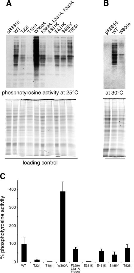
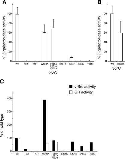
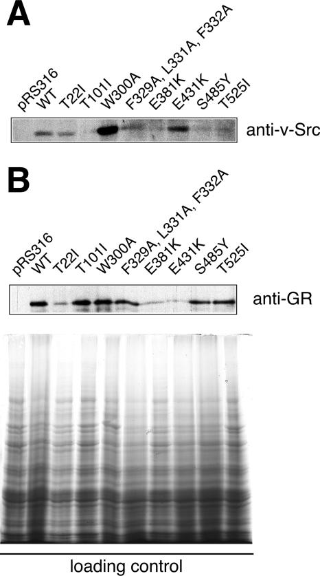
The mechanism of client protein activation by Hsp90 is enigmatic, and it is uncertain whether Hsp90 employs a common route for all proteins. Using a mutational analysis approach, we investigated the activation of two types of client proteins, glucocorticoid receptor (GR) and the kinase v-Src by the middle domain of Hsp90 (Hsp90M) in vivo. Remarkably, the overall cellular activity of v-Src was highly elevated in a W300A mutant yeast strain due to a 10-fold increase in cellular protein levels of the kinase. In contrast, the cellular activity of GR remained almost unaffected by the W300A mutation but was dramatically sensitive to S485Y and T525I exchanges. In addition, we show that mutations S485Y and T525I in Hsp90M reduce the ATP hydrolysis rate, suggesting that Hsp90 ATPase is more tightly regulated than assumed previously. Therefore, the activation of GR and v-Src has various demands on Hsp90 biochemistry and is dependent on separate functional regions of Hsp90M. Thus, Hsp90M seems to discriminate between different substrate types and to adjust the molecular chaperone for proper substrate activation.


Figures

FIG. 1.
FIG. 1.
Point mutations generated along the Hsp90 sequence and resulting phenotypes for S. cerevisiae. (A) Schematic representation of the domain organization of Hsp90. Positions of amino acid substitutions refer to the sequence of yeast Hsc82. (B) Viability of mutant yeast strains at 25 and 37°C. Shuffled cells were grown overnight in YPD medium, adjusted to 1 × 10⁸ cells per ml, and 10-fold serially diluted. Two-microliter portions were spotted onto YPD agar plates and incubated for 72 h at 25°C or for 48 h at 37°C. Boldface type indicates cells with temperature-sensitive growth defects. WT, wild type.
FIG. 2.
FIG. 2.
Interaction of p23 and Aha1 with middle domain mutants of Hsp90. Purified p23 (left panel) and Aha1 (right panel) as well as wild-type (WT) and mutant Hsp90 proteins were incubated as indicated and fractionated by gel filtration chromatography on a Superose12 column. Fractions were analyzed by SDS-polyacrylamide gel electrophoresis. Marker proteins are shown on top (thyroglobulin, 669 kDa; bovine serum albumin, 67 kDa). In the case of p23, a negative control in the absence of p23 (w/o) is included.
FIG. 3.
FIG. 3.
Quantification of Hsp90 middle domain mutants binding to Aha1 and p23 at 4, 25, and 37°C. Protein complexes of p23 or Aha1 with Hsp90 wild-type and mutant proteins were formed and fractionated by gel filtration chromatography at different temperatures as indicated. Quantification of Coomassie-stained SDS gels was performed using a Bio-Rad ChemiDoc XRS system with the Quantity One software package. Binding activity was determined as the percent ratio of bound p23 or Aha1 versus Hsp90 protein in fractions 6 to 11. One representative gel per experiment is shown. For quantification, experiments were repeated three times, and the error bars show the standard deviations for the three experiments. (A) Quantification of p23 binding to Hsp90 wild type (WT) and Hsp90 mutants. (B) Quantification of Aha1 binding to Hsp90 wild type and Hsp90 mutants.
FIG. 4.
FIG. 4.
Point mutations in the sequence of the middle domain of Hsp90 interfere with the activation of the Hsp90-dependent client proteins v-Src. (A) Cell lysates were prepared from normal or mutant ΔPCLDa/α strains expressing v-Src or from empty vector control cells (pRS316) grown at 25°C as described in Materials and Methods. Proteins were separated by polyacrylamide gel electrophoresis on 7.5% gels and transferred to nitrocellulose membranes. Phosphotyrosine activity was monitored by Western blot analysis with antibody 4G10. Quantification of protein load was performed with a Bio-Rad ChemiDoc XRS system as described in Materials and Methods and used for normalization of phosphotyrosine activity. One representative blot and gel out of three independent experiments are shown. (B) Phosphotyrosine activity of control cells (pRS316) and of wild-type and W300A cells grown at 30°C. (C) Quantification of v-Src-dependent phosphotyrosine levels from the Western blots shown in panel A. Error bars show the standard deviations from the means for the three experiments. The activity of wild-type (WT) cells was set to 100%.
FIG. 5.
FIG. 5.
GR and v-Src activity in wild-type and mutant Hsp90 yeast strains. Mutant and wild-type strains were cotransformed with GR and the reporter plasmid pSX26.1 containing β-galactosidase under the control of GR response elements. GR was activated by the addition of 10 μM DOC as described in Materials and Methods. GR-dependent β-galactosidase activity was measured using a GalactoStar kit (Tropix) and normalized to the protein concentration of the lysate. Activities are averages from at least five independent experiments, and error bars are indicated. The background activity of expressed but not DOC-activated GR was subtracted, resulting in lower specific activities than those reported in previous studies (19). (A) GR activity in wild-type and indicated mutant strains grown at 25°C. (B) GR activity in wild-type and W300A strains measured at 30°C. (C) v-Src and GR activity measured for wild-type and mutant yeast strains at 25°C. Activity of the wild-type (WT) strain was set to 100%.
FIG. 6.
FIG. 6.
Steady-state expression levels of Hsp90-dependent client proteins v-Src and GR in wild-type and mutant yeast strains. Quantification was performed with a Bio-Rad ChemiDoc XRS system using the Quantity One software package and normalized to the protein content of loading controls. A typical blot for v-Src and GR levels is shown, and experiments were done in triplicate for quantification. Data were used to calculate specific activity of v-Src and GR as presented in Table 2. (A) Cell lysates from v-Src expressing yeast (same as in Fig. 4A, see that figure legend for loading control) were blotted for v-Src protein levels using the specific monoclonal antibody EC10. (B) Cell lysates from GR-expressing yeast were blotted for GR protein levels using the specific monoclonal antibody BuGR2. Loading controls are shown on the bottom. WT, wild type.
FIG. 7.
FIG. 7.
Effect of cochaperone overexpression on the viability and client protein activity of Hsp90 mutant strains S485Y and T525I. (A) S485Y and T525I strains were cotransformed with empty vector serving as a control or p23, Hop, Aha1, and p50 expression plasmids. Pictures were taken from different plates that had been incubated together at 37°C. (B) GR activity measured for wild-type and mutant yeast strains cotransformed with empty vector, p23, Hop, Aha1, or p50 expression plasmids. Activity of the wild-type (WT) strain was set to 100%.

Similar articles

Cited by

References

    1. Ali, M. M. U., S. M. Roe, C. K. Vaughan, P. Meyer, B. Panaretou, P. W. Piper, C. Prodromou, and L. H. Pearl. 2006. Crystal structure of an Hsp90-nucleotide-p23/Sba1 closed chaperone complex. Nature 440:1013-1017. - PMC - PubMed
    1. Bohen, S. P., and K. R. Yamamoto. 1993. Isolation of Hsp90 mutants by screening for decreased steroid receptor function. Proc. Natl. Acad. Sci. USA 90:11424-11428. - PMC - PubMed
    1. Brychzy, A., T. Rein, K. F. Winklhofer, F. U. Hartl, J. C. Young, and W. M. J. Obermann. 2003. Cofactor Tpr2 combines two TPR domains and a J domain to regulate the Hsp70/Hsp90 chaperone system. EMBO J. 22:3613-3623. - PMC - PubMed
    1. Dittmar, K. D., D. R. Demady, L. F. Stancato, P. Krishan, and W. B. Pratt. 1997. Folding of the glucocorticoid receptor by heat shock protein (hsp) 90-based chaperone machinery. J. Biol. Chem. 272:21213-21220. - PubMed
    1. Fang, Y. F., A. E. Fliss, J. Rao, and A. J. Caplan. 1998. Sba1 encodes a yeast Hsp90 cochaperone that is homologous to vertebrate p23 proteins. Mol. Cell. Biol. 18:3727-3734. - PMC - PubMed

Publication types

MeSH terms

LinkOut - more resources