. 2006 Sep 25;174(7):997-1007.
doi: 10.1083/jcb.200605004. Epub 2006 Sep 18.

Ca2+ and synaptotagmin VII-dependent delivery of lysosomal membrane to nascent phagosomes

Affiliations

Ca2+ and synaptotagmin VII-dependent delivery of lysosomal membrane to nascent phagosomes

Cecilia Czibener et al. J Cell Biol. .

Abstract

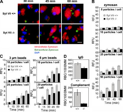
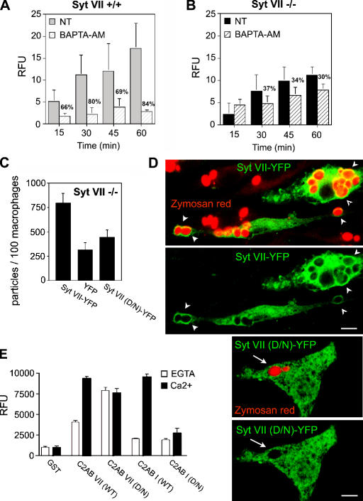
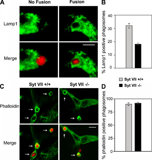
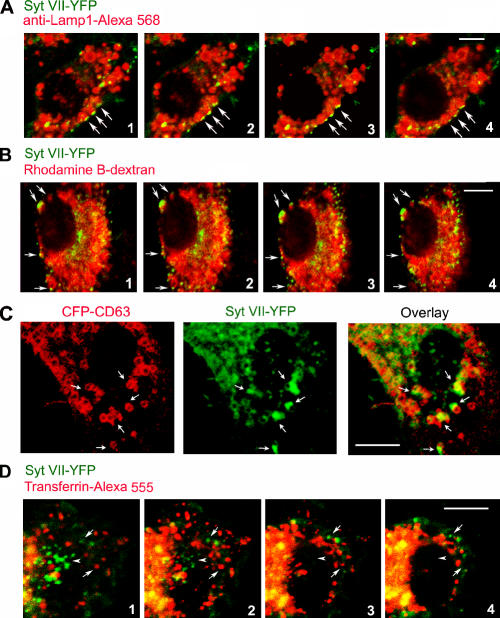
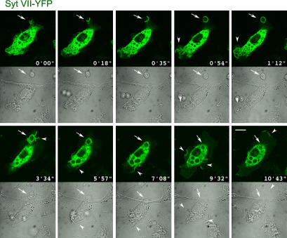
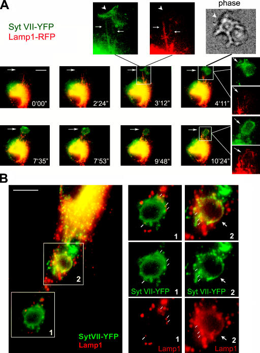
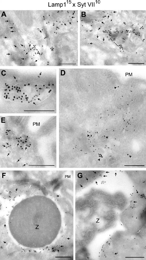
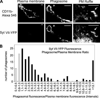
Synaptotagmin (Syt) VII is a ubiquitously expressed member of the Syt family of Ca2+ sensors. It is present on lysosomes in several cell types, where it regulates Ca2+-dependent exocytosis. Because [Ca2+]i and exocytosis have been associated with phagocytosis, we investigated the phagocytic ability of macrophages from Syt VII-/- mice. Syt VII-/- macrophages phagocytose normally at low particle/cell ratios but show a progressive inhibition in particle uptake under high load conditions. Complementation with Syt VII rescues this phenotype, but only when functional Ca2+-binding sites are retained. Reinforcing a role for Syt VII in Ca2+-dependent phagocytosis, particle uptake in Syt VII-/- macrophages is significantly less dependent on [Ca2+]i. Syt VII is concentrated on peripheral domains of lysosomal compartments, from where it is recruited to nascent phagosomes. Syt VII recruitment is rapidly followed by the delivery of Lamp1 to phagosomes, a process that is inhibited in Syt VII-/- macrophages. Thus, Syt VII regulates the Ca2+-dependent mobilization of lysosomes as a supplemental source of membrane during phagocytosis.


Figures

Figure 1.
Figure 1.
The phagocytosis defect of Syt VII−/− BMMs increases with particle load and size. (A) Syt VII+/+ or VII−/− BMMs were exposed to 25 IgG-opsonized zymosan red particles/cell for the periods of time indicated. Cells were washed, fixed, and stained without permeabilization to detect extracellular particles (green) and nuclei (DAPI, blue). Bar, 10 μm. (B) The phagocytosis defect of Syt VII−/− BMMs increases with particle load. Opsonized zymosan red uptake was determined fluorometrically at the indicated time points after incubation with increasing numbers of particles/cell. (C) The phagocytosis defect of Syt VII−/− BMMs increases with particle size. The uptake of IgG-opsonized fluorescent polystyrene particles was determined fluorometrically at the indicated time points after incubation with 10 or 25 particles/cell. RFU, relative fluorescence unit. (D) The uptake defect of Syt VII−/− BMMs is independent of the receptor-mediating phagocytosis. The number of internalized sheep RBCs opsonized with IgG or complement was determined after BMM exposure to 10 particles/cell for 30 min. The data represent the mean ± SD (error bars) of triplicates.
Figure 2.
Figure 2.
Ca2+-dependent Syt VII activity regulates phagocytosis. (A and B) Residual phagocytosis of Syt VII−/− BMMs is less dependent on [Ca2+]i. BMMs pretreated or not treated (NT) with BAPTA-AM were exposed to 10 IgG-opsonized zymosan red/cell. After the indicated time points, cells were washed, fixed, and the number of internalized particles were determined fluorometrically. (A) Syt VII+/+ BMMs. (B) Syt VII−/− BMMs. Percent inhibition for each time point is indicated. (C) Syt VII–YFP restores phagocytosis to wild-type levels. Syt VII−/− BMMs were transduced with Syt VII–YFP, YFP, or Syt VII (D/N)–YFP (full-length Syt VII with mutations on the four putative Ca2+-binding residues) and exposed to 25 IgG-opsonized zymosan red/cell for 1 h. (D) Syt VII (D/N)–YFP is not recruited to phagosomes as efficiently as Syt VII–YFP. Confocal images of the BMMs in C, acquired under identical conditions. Both constructs were targeted to tubular intracellular compartments, but only Syt VII–YFP was efficiently recruited to zymosan red–containing phagosomes (arrowheads, top). Arrows in the bottom panels point to Syt VII (D/N)–YFP-negative phagosomes. The YFP signal was enhanced by immunofluorescence with anti-GFP antibodies. Bars, 5 μm. (E) Mutations in the predicted Ca2+-binding residues shift the PS binding of C2AB VII from Ca2+ dependent to Ca2+ independent. The binding of GST–C2AB VII, GST–C2AB VII (D/N), GST–C2AB I, and GST–C2AB I (D/N) to PS-containing liposomes was determined. Proteins were immobilized on glutathione–Sepharose beads and assayed for the binding of PS/PC liposomes in the presence of 2 mM EGTA (white bars) or 0.2 mM Ca2+ (black bars). RFU, relative fluorescence unit. The data represent the mean ± SD (error bars) of triplicates.
Figure 3.
Figure 3.
The early fusion of lysosomes with phagosomes is impaired in Syt VII−/− BMMs. BMMs were exposed to opsonized zymosan red for 10 min, fixed, and stained with anti-Lamp1 mAbs (A and B) or phalloidin (C and D). (A) Examples of the morphological criteria used to score phagosomes for fusion or no fusion. (B) Percentage of phagosomes that fused with Lamp1-containing lysosomes. (C) Images of phalloidin-stained recently formed phagosomes in Syt VII+/+ or VI−/− BMMs. The arrows point to phagosomes containing zymosan red surrounded by phalloidin-stained polymerized actin. (D) Percentage of phalloidin-positive phagosomes. (B and D) The data represent the mean ± SD (error bars) of triplicates. Bars, 5 μm.
Figure 4.
Figure 4.
Syt VII–YFP colocalizes with peripheral domains of late endosomes/lysosomes. (A) BMMs were transduced with Syt VII–YFP, fixed, and stained with anti-Lamp1 mAbs. (B) BMMs were transduced with Syt VII–YFP, and the lysosomal compartment was loaded with rhodamine B–dextran before fixation. (A and B) Images are sequential 0.8-μm optical slices of a confocal z stack. (C) BMMs were transduced with Syt VII–YFP and CFP-CD63 and examined by live confocal microscopy. (A–C) The arrows point to domains enriched in Syt VII–YFP that were closely associated with Lamp1-positive compartments (A), rhodamine B–dextran-containing compartments (B), or compartments containing CFP-CD63 (C). (D) BMMs were transduced with Syt VII–YFP, and early endosomes were loaded with AlexaFluor555-transferrin before fixation. Images are sequential 0.8-μm optical slices of a confocal z stack. Arrows point to compartments containing Syt VII–YFP or AlexaFluor555-transferrin that are not in close association with each other. Arrowheads point to a cluster of Syt VII–enriched domains that are not associated with transferrin-containing early endosomes. Bars, 5 μm.
Figure 5.
Figure 5.
Syt VII–YFP is rapidly recruited to phagocytic cups and to nascent phagosomes. Selected frames from a confocal time-lapse video of a BMM transduced with Syt VII–YFP and exposed to 10 IgG-opsonized zymosan particles/cell (Video 1, available at http://www.jcb.org/cgi/content/full/jcb.200605004/DC1). The images show the rapid recruitment of Syt VII–YFP to sites of particle attachment and to expanding phagocytic cups (arrows). At later time points, Syt VII–YFP recruitment is also detected on areas of plasma membrane ruffling, which were associated (frames at 1 min 12 s and at 3 min 34 s) or not associated (frames at 9 min 32 s and at 10 min 43 s) with particle uptake (arrowheads). Top panels, YFP channel; bottom panels, differential interference contrast. Bar, 10 μm.
Figure 6.
Figure 6.
Syt VII–YFP and Lamp1-RFP are sequentially delivered from tubular lysosomes to nascent phagosomes. (A) Selected frames from a wide-field time-lapse video of a BMM transduced with Syt VII–YFP and Lamp1-RFP and exposed to 10 IgG-opsonized zymosan particles/cell (Videos 2–4, available at http://www.jcb.org/cgi/content/full/jcb.200605004/DC1). Frames from 0 min 0 s to 3 min 12 s: initial colocalization of Syt VII–YFP and Lamp1-RFP on tubular lysosomes expanding toward a site of phagosome formation. The arrow on the merged image at 3 min 12 s points to the boxed area reproduced in the enlarged, dissociated images above. Arrows point to tubular lysosomes containing both markers, and arrowheads point to Syt VII–YFP recruited to phagocytic cups (phase-contrast image, top). In the subsequent time points, Lamp1-containing tubules are seen wrapping themselves around Syt VII–YFP-containing phagosomes (frames from 7 min 35 s to 10 min 24 s; Videos 2–4). Dissociated, enlarged images are indicated by white lines originating from the merged image. Arrows point to the site of zymosan uptake. (B) Confocal microscopy images of BMMs transduced with Syt VII–YFP, exposed to IgG-opsonized zymosan for 20 min, fixed, and stained with anti-Lamp1 mAbs (red). In the same BMMs, two patterns of Lamp1 association with phagosomes can be seen (right). (1) Lamp1 patches (white lines) in close association with Syt VII–YFP-containing phagosomes (patches are likely to correspond to Lamp1 tubules surrounding the phagosome; A). (2) Lamp1 and Syt VII–YFP fully colocalize on the phagosome membrane (arrows). Bars (A), 10 μm; (B) 5 μm.
Figure 7.
Figure 7.
Syt VII and Lamp1 are localized on distinct microdomains of tubulovesicular lysosomes and phagosomes. (A–C) Immunogold labeling of BMM ultrathin cryosections detected endogenous Syt VII and Lamp1 on apparently contiguous microdomains of tubulovesicular compartments. 15 nm gold, mAbs to Lamp1; 10 nm gold, antibodies to Syt VII. (D and E) Immunogold labeling of Syt VII–YFP in transduced BMMs showing that its distribution is similar to the endogenous protein (associated with Lamp1 on peripheral tubulovesicular compartments). 15 nm gold, mAbs to Lamp1; 10 nm gold, anti-GFP antibodies. In A, B, C, and E, the arrows point to gold particles labeling Syt VII; in D, the arrowheads point to gold particles labeling Lamp1. (F and G) Immunogold detection of endogenous Syt VII and Lamp1 on microdomains of recently formed phagosomes (BMMs were incubated with IgG-opsonized zymosan for 10 min). In F, the ruffling plasma membrane is visible on the top right (PM). Arrows point to gold particles detecting Syt VII on phagosome-associated membranes. 15 nm gold, mAbs to Lamp1; 10 nm gold, antibodies to Syt VII. Z, zymosan particle. Bars (A–E), 200 nm; (F and G) 400 nm.
Figure 8.
Figure 8.
Syt VII–YFP-containing intracellular compartments contribute to phagosome formation. (A) Representative confocal images of anti-CD11b–AlexaFluor546 antibodies or Syt VII–YFP on the plasma membrane, phagosomes (arrowheads), and plasma membrane ruffles (arrows) of transduced BMMs. Bars, 5 μm. (B) Quantitation of the relative fluorescence intensity of Syt VII–YFP on 206 phagosomes. Each bar represents the number of phagosomes with a phagosome/plasma membrane fluorescence ratio within the intervals indicated. Black bars, phagosomes (81% of the total) in which Syt VII–YFP was enriched in relation to the plasma membrane (ratio of >1). White bars, phagosomes showing a ratio of <1. Data were normalized to the fluorescence intensity of the plasma membrane of the same cell.

Similar articles

Cited by

References

    1. Aderem, A., and D.M. Underhill. 1999. Mechanisms of phagocytosis in macrophages. Annu. Rev. Immunol. 17:593–623. - PubMed
    1. Advani, R.J., H.R. Bae, J.B. Bock, D.S. Chao, Y.C. Doung, R. Prekeris, J.S. Yoo, and R.H. Scheller. 1998. Seven novel mammalian SNARE proteins localize to distinct membrane compartments. J. Biol. Chem. 273:10317–10324. - PubMed
    1. Advani, R.J., B. Yang, R. Prekeris, K.C. Lee, J. Klumperman, and R.H. Scheller. 1999. VAMP-7 mediates vesicular transport from endosomes to lysosomes. J. Cell Biol. 146:765–776. - PMC - PubMed
    1. Arantes, R.M., and N.W. Andrews. 2006. A role for synaptotagmin VII-regulated exocytosis of lysosomes in neurite outgrowth from primary sympathetic neurons. J. Neurosci. 26:4630–4637. - PMC - PubMed
    1. Bajno, L., X.R. Peng, A.D. Schreiber, H.P. Moore, W.S. Trimble, and S. Grinstein. 2000. Focal exocytosis of VAMP3-containing vesicles at sites of phagosome formation. J. Cell Biol. 149:697–706. - PMC - PubMed

Publication types