. 2006 Sep 25;174(7):1059-69.
doi: 10.1083/jcb.200605113. Epub 2006 Sep 18.

HDAC3 is crucial in shear- and VEGF-induced stem cell differentiation toward endothelial cells

Affiliations

HDAC3 is crucial in shear- and VEGF-induced stem cell differentiation toward endothelial cells

Lingfang Zeng et al. J Cell Biol. .

Abstract

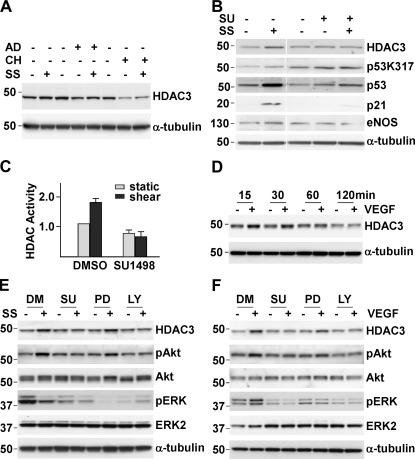
Reendothelialization involves endothelial progenitor cell (EPC) homing, proliferation, and differentiation, which may be influenced by fluid shear stress and local flow pattern. This study aims to elucidate the role of laminar flow on embryonic stem (ES) cell differentiation and the underlying mechanism. We demonstrated that laminar flow enhanced ES cell-derived progenitor cell proliferation and differentiation into endothelial cells (ECs). Laminar flow stabilized and activated histone deacetylase 3 (HDAC3) through the Flk-1-PI3K-Akt pathway, which in turn deacetylated p53, leading to p21 activation. A similar signal pathway was detected in vascular endothelial growth factor-induced EC differentiation. HDAC3 and p21 were detected in blood vessels during embryogenesis. Local transfer of ES cell-derived EPC incorporated into injured femoral artery and reduced neointima formation in a mouse model. These data suggest that shear stress is a key regulator for stem cell differentiation into EC, especially in EPC differentiation, which can be used for vascular repair, and that the Flk-1-PI3K-Akt-HDAC3-p53-p21 pathway is crucial in such a process.


Figures

Figure 1.
Figure 1.
Laminar flow resulted in ES cell–derived progenitor cell proliferation and differentiation into ECs. (A) Shear-increased ES and Sca1+ cell proliferation. (B) Shear-increased Flk-1 and eNOS protein levels in ES cells. ES, routinely cultured ES cell; ST, static; SH, shear. (C) FACS analysis for EC marker expression, including CD31, CD133, and CD106, in response to shear stress for 24 h. *, P < 0.05. (D) In vitro tube-formation assay with static and sheared-ES cells on Matrigel. (E) Hematoxylin and eosin (HE) and X-gal staining for sections from in vivo angiogenesis assay at day 14. The data are representatives or means ± the SD of three independent experiments. Bars, 100 μm.
Figure 2.
Figure 2.
Up-regulation of p53 and p21 correlated with shear-induced EC differentiation. (A) Shear up-regulated p53 and p21 proteins in ES cells. SS, shear stress. (B) Double immunofluorescence staining showing the colocalization of p53 or p21 with CD106 in sheared-ES cells. (C) Overexpression of p53 and p21 increased EC marker reporter gene expression in ES cells. (D) FACS analysis showing the effect of overexpression of p53 by Ad-p53 (multiplicity of infection [MOI] = 50) on CD31, CD133, CD106, and CD144 expression. Ad-LacZ virus was included as a control. *, P < 0.05. (E) Representative images showing X-gal, anti-p53, p21, and CD144 staining of serial sections of inoculates from Ad-p53–infected ES cells. The data are representatives or means ± the SD from three independent experiments. Bars, 50 μm.
Figure 3.
Figure 3.
Knockdown of p53 and p21 ablated shear-induced EC differentiation. (A) Western blot analysis showing the effect of p53-siRNA on shear-induced p53, p21, eNOS, and Flt-1 proteins. Untransfected cells (ctl) and control siRNA (ctlsi) were included as controls. (B) Western blot analysis showing the effect of p21 siRNA on shear-induced p21 expression, eNOS, Flk-1, and Flt-1 protein production. The data are representative of three independent experiments.
Figure 4.
Figure 4.
HDAC3 contributed to shear-induced p53 deacetylation and p21 activation. (A) Western blot to detect p53 acetylation status at Lys317 and Lys370 and p21 activation in duplicate slides from cells sheared for 24 h. (B) HDAC activity detected in static and shear samples harvested at the times indicated. (C) HDAC1 and HDAC3 proteins were up-regulated by shear treatment. (D) Overexpression of HDAC3 enhanced p53-mediated p21 reporter gene expression. The data are means ± the SD of three independent experiments.
Figure 5.
Figure 5.
Inhibition of HDAC3 ablated shear-induced p53 deacetylation, p21 activation, and EC differentiation. (A) TSA abolished shear-induced HDAC activation. (B) TSA blocked shear-induced HDAC3 activation, p53 deacetylation, p21 activation, and EC differentiation. (C) Western blot data showing the effect of HDAC3 siRNA on shear-induced p53, acetyl-p53Lys370, p21, HDAC3, and Flt-1 proteins. Untransfected cells (ctl) and control siRNA (ctlsi) were included as controls. (D) Overexpression of p21 partially rescued HDAC3 siRNA–mediated suppression of Flt-1 reporter gene expression. The data are representatives or means ± the SD from three independent experiments.
Figure 6.
Figure 6.
Shear- and VEGF-stabilized HDAC3 through Flk-1–PI3K–Akt signal pathways. (A) RNA and protein synthesis inhibitors did not affect shear-increased HDAC3 protein production. (B) Flk-1 inhibitor ablated shear-induced HDAC3 stabilization, p53 deacetylation, p21 activation, and EC differentiation. (C) Flk-1 inhibitor abolished shear-induced HDAC activation. (D) VEGF transiently up-regulated HDAC3 protein induction. (E) Western blot data showed that shear-induced HDAC3 stabilization was related to Flk-1–mediated Akt phosphorylation. (F) Western blot analysis revealed that VEGF-induced HDAC3 stabilization was related to Flk-1–mediated Akt phosphorylation. AD, actinomycin D; CH, cycloheximide; LY, LY294002; PD, PD98059; SS, shear stress; SU, SU1498. The data are representatives or means ± the SD from three independent experiments.
Figure 7.
Figure 7.
HDAC3 was involved in VEGF-induced EC differentiation and blood vessel formation in vivo. (A) HDAC3 siRNA ablated VEGF-induced EC marker expression in ES cells. The data are means ± the SD of three independent experiments. (B) A representative of immunohistological staining for CD144, HDAC3, p53, and p21 in blood vessels formed by VEGF-treated Sca1+ cells. (C) A representative of the X-gal staining and immunohistological staining for CD31, HDAC3, and p21 in blood vessels in D13.5 embryonic sections from Tie2-LacZ/ApoE−/− mice. Bars, 200 μm.
Figure 8.
Figure 8.
Sca1+ cells incorporated into injured artery and reduced lesion formation in ApoE−/− mice. (A) X-gal and double immunofluorescence staining showed the incorporation of VEGF-treated Sca1+ cells into the injured femoral artery. (B) VEGF-treated Sca1+ cell reduced the lesion formation caused by artery injury. a, uninjured artery; b, injured artery without cell treatment; c, injured artery with cell treatment; d, higher magnification of image c; e, statistical analysis of the ratio of neointima to the media. (C) Shear stress–induced Sca1+ cell reduced the lesion formation caused by artery injury. a, uninjured artery; b, injured artery without cell treatment; c, X-gal staining showed the incorporated Sca1+ cell; d, injured artery with Sca1+ cell treatment; e, statistical analysis of the ratio of neointima to the media. Data are means ± the SD from six animals per group. Bars: (A–C) 100 μm; (c) 50 μm.

Similar articles

Cited by

References

    1. Akimoto, S., M. Mitsumata, T. Sasaguri, and Y. Yoshida. 2000. Laminar shear stress inhibits vascular endothelial cell proliferation by inducing cyclin-dependent kinase inhibitor p21(Sdi1/Cip1/Waf1). Circ. Res. 86:185–190. - PubMed
    1. Bruhl, T., C. Heeschen, A. Aicher, A.S. Jadidi, J. Haendeler, J. Hoffmann, M.D. Schneider, A.M. Zeiher, S. Dimmeler, and L. Rossig. 2004. p21Cip1 levels differentially regulate turnover of mature endothelial cells, endothelial progenitor cells, and in vivo neovascularization. Circ. Res. 94:686–692. - PubMed
    1. Byrne, A.M., D.J. Bouchier-Hayes, and J.H. Harmey. 2005. Angiogenic and cell survival functions of vascular endothelial growth factor (VEGF). J. Cell. Mol. Med. 9:777–794. - PMC - PubMed
    1. Claesson-Welsh, L. 2003. Signal transduction by vascular endothelial growth factor receptors. Biochem. Soc. Trans. 31:20–24. - PubMed
    1. Coultas, L., K. Chawengsaksophak, and J. Rossant. 2005. Endothelial cells and VEGF in vascular development. Nature. 438:937–945. - PubMed

Publication types

MeSH terms